1
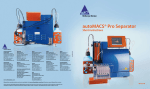
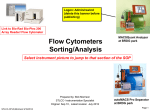
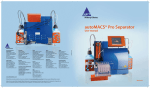
CD3+CD56+ NKT Cell
Isolation Kit
human
Order no. 130-093-064
Index
1.1 Principle of MACS® Separation
1. Description
The CD3+CD56+ NKT Cell Isolation Kit is a two-step magnetic
labeling system for the isolation of CD3+CD56+ NKT cells from human
peripheral blood mononuclear cells (PBMCs). In the first step, NK cells
and monocytes are indirectly magnetically labeled by using a cocktail of
biotin-conjugated antibodies and Anti-Biotin MicroBeads. The
labeled cells are subsequently depleted by separation over a MACS®
Column.
In the second step, CD3+CD56+ NKT cells are directly labeled
with CD56 MicroBeads and isolated by positive selection from
the pre-enriched NKT cell fraction. The magnetically labeled
CD3+CD56+ NKT cells are retained on the column and eluted after
removal of the column from the magnetic field.
1.1 Principle of MACS® Separation
1.2 Background and product applications
1.3 Reagent and instrument requirements
2. Protocol
2.1 Sample preparation
2.2 Magnetic labeling of non-CD3+CD56+ NKT cells
2.3 Magnetic separation:
Depletion of non-CD3+CD56+ NKT cells
2.4 Magnetic labeling of CD3+CD56+ NKT cells
2.5 Magnetic separation:
Positive selection of CD3+CD56+ NKT cells
2.6 Evaluation of CD3+CD56+ NKT cell purity
3. Example of a separation using the CD3+CD56+ NKT Cell
Isolation Kit
4. References
1. Description
Components
2 mL CD3+CD56+ NKT Biotin-Antibody
Cocktail, human:
Cocktail of biotin-conjugated monoclonal
anti-human antibodies against antigens not
expressed by CD3+CD56+ NKT cells.
2 mL Anti-Biotin MicroBeads:
MicroBeads conjugated to monoclonal antibiotin antibody (isotype: mouse IgG1).
Human PBMCs
Depletion of non-CD3+CD56+ NKT cells
1. Indirect magnetic labeling of nonCD3+CD56+ NKT cells with BiotinAntibody Cocktail and Anti-Biotin
MicroBeads.
2. Magnetic separation using LD Column
or autoMACS (programs "Depl05" or
Flow-through fraction:
"Depl025").
pre-enriched CD3+CD56+ NKT
cells
Positive selection of CD3+CD56+ NKT cells
1. Direct magnetic labeling of CD3+CD56+ NKT
cells with CD56 MicroBeads.
2. Magnetic separation using an MS Column or
autoMACS (program "Possel").
2 mL CD56 MicroBeads:
MicroBeads conjugated to monoclonal antiCD56 antibody (clone AF12-7H3; isotype:
mouse IgG1).
Size
For 2×10⁹ total cells, up to 20 separations.
Product format
All components are supplied in buffer containing
stabilizer and 0.05% sodium azide.
Storage
Store protected from light at 2–8 °C. Do not freeze.
The expiration date is indicated on the vial label.
Elution from column:
CD3+CD56+ NKT cells
1.2 Background and product applications
Natural killer (NK) T cells represent a subpopulation of T cells
that possess properties of NK cells.¹,² NKT cells can be stimulated
through contact with antigen or by cytokines such as IL-12 to release
large amounts of cytokines and to exert cytotoxic effects. NKT cells
are a crucial part of the innate immune system³ and have an influence
on the development of autoimmune diseases. They are also involved
in tumor immunology as well as immunity against viruses, bacterial⁴,
fungal, and parasitic pathogens.
Example applications
140-001-942.03
●
Cytokine production analysis after activation/stimulation.
●
Gene expression analysis of NKT cell subsets.
●
Analysis of the functional role of NKT cell surface receptors.
●
Studies on cytotoxic and cytolytic activity.
www.miltenyibiotec.com
Miltenyi Biotec GmbH
Friedrich-Ebert-Str. 68
51429 Bergisch Gladbach, Germany
Phone +49-2204-8306-0 Fax +49-2204-85197
Miltenyi Biotec Inc.
12740 Earhart Avenue, Auburn CA 95602, USA
Phone 800 FOR MACS, 530 888-8871 Fax 530 888-8925
page 1/4
Order no. 130-093-064
●
Generation of NKT cell lines.
2. Protocol
●
Interaction with dendritic cells.
2.1 Sample preparation
1.3 Reagent and instrument requirements
●
Buffer: Prepare a solution containing phosphate-buffered saline
(PBS) pH 7.2, 0.5% bovine serum albumin (BSA), and 2 mM
EDTA by diluting MACS® BSA Stock Solution (# 130-091-376)
1:20 with autoMACS™ Rinsing Solution (# 130-091-222). Keep
buffer cold (4−8 °C). Degas buffer before use, as air bubbles could
block the column.
▲ Note: EDTA can be replaced by other supplements such as anticoagulant citrate
dextrose formula-A (ACD-A) or citrate phosphate dextrose (CPD). BSA can be
replaced by other proteins such as human serum albumin, human serum, or fetal calf
serum. Buffers or media containing Ca2+ or Mg2+ are not recommended for use.
● MACS Columns and MACS Separators: Depletion of nonCD3+CD56+ NKT cells is performed on an LD Column. The
subsequent positive selection of CD3+CD56+ NKT cells is
performed on an MS Column. Depletion and positive selection
can also be performed by using the autoMACS Separator.
Column max. number
of labeled cells
5 ×108
LD
MidiMACS, QuadroMACS,
VarioMACS, SuperMACS
Positive selection
2×10⁸
MS
107
MiniMACS, OctoMACS,
VarioMACS, SuperMACS
Depletion and positive selection
autoMACS
2 ×108
4 ×109
autoMACS
▲ Note: Column adapters are required to insert certain columns into VarioMACS™
Separator or SuperMACS™ Separator. For details, see MACS Separator data sheets.
●
●
▲ Note: To remove platelets after density gradient separation, resuspend cell pellet
in buffer and centrifuge at 200×g for 10−15 minutes at 20 °C. Carefully aspirate
supernatant. Repeat washing step.
When working with tissues, prepare a single-cell suspension by
a standard preparation method. For details see section General
Protocols in the User Manuals or visit www.miltenyibiotec.com/
protocols.
▲ Note: Dead cells may bind non-specifically to MACS MicroBeads. To remove dead
cells, we recommend using density gradient centrifugation or the Dead Cell Removal
Kit (# 130-090-101).
2.2
Magnetic labeling of non-CD3+CD56+ NKT cells
max. number Separator
of total cells
Depletion
108
When working with anticoagulated peripheral blood or
buffy coat, PBMCs should be isolated by density gradient
centrifugation, e.g. using Ficoll-Paque™. For details see
section General Protocols in the User Manuals or visit
www.miltenyibiotec.com/protocols.
(Optional) Fluorochrome-conjugated antibody for flow
cytometric analysis, e.g. CD3-FITC (# 130-080-401) and CD56PE (BD™ Biosciences, NCAM16.2).
(Optional) Propidium iodide (PI) or 7-AAD for flow cytometric
exclusion of dead cells without cell fixation. For cell fixation and
flow cytometric exclusion of dead cells, the Fixation and Dead
Cell Discrimination Kit (# 130-091-163) is recommended.
● (Optional) Pre-Separation Filters (# 130-041-407) to remove cell
clumps.
▲ Work fast, keep cells cold, and use pre-cooled solutions. This will
prevent capping of antibodies on the cell surface and non-specific
cell labeling.
▲ Volumes for magnetic labeling given below are for up to
10⁸ total cells. When working with fewer than 10⁸ cells, use the same
volumes as indicated. When working with higher cell numbers, scale
up all reagent volumes and total volumes accordingly (e.g. for 2×10⁸
total cells, use twice the volume of all indicated reagent volumes and
total volumes).
▲ For optimal performance it is important to obtain a single‑cell
suspension before magnetic separation. Pass cells through 30 µm
nylon mesh (Pre-Separation Filters, # 130-041-407) to remove cell
clumps which may clog the column.
1.
Determine cell number.
2. Centrifuge cell suspension at 300×g for 10 minutes. Aspirate
supernatant completely.
3. Resuspend cell pellet in 400 µL of buffer per 10⁸ cells.
4. Add 100 µL of CD3+CD56+ NKT Cell Biotin-Antibody
Cocktail per 10⁸ cells.
5. Mix well and refrigerate for 10 minutes (4–8 °C).
6. Wash cells by adding 5–10 mL of buffer and centrifuge at 300×g
for 10 minutes. Aspirate supernatant completely.
7. Resuspend cell pellet in 400 µL of buffer per 10⁸ cells.
8. Add 100 µL of Anti-Biotin MicroBeads per 10⁸ cells.
9. Mix well and refrigerate for 15 minutes (4–8 °C).
10. Wash cells by adding 5–10 mL of buffer and centrifuge at 300×g
for 10 minutes. Aspirate supernatant completely.
11. Resuspend up to 108 cells in 500 µL of buffer.
▲ Note: For larger cell numbers, scale up buffer volume accordingly.
140-000-942.03
12. Proceed to magnetic separation (2.3).
www.miltenyibiotec.com
Unless otherwise specifically indicated, all Miltenyi Biotec
products and services are for research use only and not for
diagnostic or therapeutic use.
page 2/4
Order no. 130-093-064
2.3 Magnetic separation: Depletion of non-CD3+CD56+
NKT cells
2.5 Magnetic separation: Positive selection of
CD3+CD56+ NKT cells
Depletion with LD Column
1.
Place LD Column in the magnetic field of a suitable MACS®
Separator. For details see LD Column data sheet.
2. Prepare column by rinsing with 2 mL of buffer.
Positive selection with MS Columns
1.
Place MS Column in the magnetic field of a suitable MACS
Separator. For details see MS Column data sheet.
3. Apply cell suspension onto the column.
2. Prepare column by rinsing with 500 µL of buffer.
4. Collect unlabeled cells which pass through and wash column
with 2×1 mL of buffer. Perform washing steps by adding buffer
successively once the column reservoir is empty. Collect total
effluent. This contains the unlabeled pre-enriched CD3+CD56+
NKT cell fraction.
3. Apply cell suspension onto the column.
5. Proceed to 2.4 for the isolation of CD3+CD56+ NKT cells.
Depletion with the autoMACS™ Separator
▲ Refer to the "autoMACS™ User Manual" for instructions on how to
use the autoMACS Separator.
1.
4. Collect unlabeled cells which pass through and wash column
with 3×500 µL of buffer. Perform washing steps by adding buffer
three times. Only add new buffer when the column reservoir is
empty.
5. Remove column from the separator and place it on a suitable
collection tube.
6. Pipette 1 mL of buffer onto the column. Immediately flush out
the fraction with magnetically labeled CD3+CD56+ NKT cells
by firmly pushing the plunger into the column.
Prepare and prime autoMACS Separator.
Positive selection with the autoMACS Separator
2. Place tube containing the magnetically labeled cells in the
autoMACS Separator. Choose separation program "Depl05". If
high purity of the CD3+CD56+ NKT cell population is desired or
if the frequency of CD3+CD56+ NKT cells in the sample is below
5%, it is recommended to use "Depl025".
▲ Refer to the "autoMACS User Manual" for instructions on how to
use the autoMACS Separator.
3. Collect unlabeled fraction from outlet port neg1. This is the preenriched CD3+CD56+ NKT cell fraction.
2. Place tube containing the magnetically labeled cells in the
autoMACS Separator. Choose separation program "Possel".
4. Proceed to 2.4 for the enrichment of CD3+CD56+ NKT cells.
3. Collect positive fraction from outlet port pos1. This is the
enriched CD3+CD56+ NKT cell fraction.
2.4 Magnetic labeling of CD3+CD56+ NKT cells
▲ Volumes for magnetic labeling given below are for an initial starting
cell number of up to 10⁸ cells. For larger initial cell numbers, scale up
volumes accordingly.
1.
Centrifuge cells at 300×g for 10 minutes. Aspirate supernatant
completely.
1.
Prepare and prime autoMACS Separator.
2.6 (Optional) Evaluation of CD3+CD56+ NKT cell purity
The purity of the enriched CD3+CD56+ NKT cells or any intermediate
fraction can be evaluated by flow cytometry or fluorescence
microscopy. Stain aliquots of the cell fractions with fluorochromeconjugated antibodies against CD3, e.g., CD3-FITC (# 130-080-401)
and against CD56, e.g., CD56-PE (BD™ Biosciences, NCAM16.2)
according to the manufacturer’s recommendations.
2. Resuspend cell pellet in 400 µL of buffer per 10⁸ initial cells.
3. Add 100 µL of CD56 MicroBeads per 10⁸ initial cells.
4. Mix well and refrigerate for 15 minutes (4–8 °C).
5. Wash cells by adding 5–10 mL of buffer and centrifuge at 300×g
for 10 minutes. Aspirate supernatant completely.
6. Resuspend up to 108 cells in 500 µL of buffer.
7. Proceed to magnetic separation (2.5).
140-001-942.03
www.miltenyibiotec.com
Unless otherwise specifically indicated, all Miltenyi Biotec
products and services are for research use only and not for
diagnostic or therapeutic use.
page 3/4
Order no. 130-093-064
3. Example of a separation using the CD3+CD56+
NKT Cell Isolation Kit
CD3+CD56+ NKT cells were isolated from human PBMCs by using
the CD3+CD56+ NKT Cell Isolation Kit, an LD and an MS Column,
a MidiMACS™ Separator and a MiniMACS™ Separator. The cells were
fluorescently stained with CD3-FITC and CD56-APC. Cell debris
and dead cells were excluded from the analysis based on scatter
signals and PI fluorescence.
Before separation
4. References
1.
Jiang, H. and Chess, L. (2006) Regulation of immune responses by T cells. N. Engl.
J. Med. 354: 1166–1176.
2.
Linsen, L. et al. (2006) Immunoregulation of autoimmunity by natural killer T cells.
Hum. Immunol. 12: 1193–1202.
3.
Carnaud, C. et al. (1999) Cutting edge: cross-talk between cells of the innate
immune system: NKT cells rapidly activate NK cells. J. Immunol. 163: 4647–4650.
4.
Bilenki, L. et al. (2005) NKT cell activation promotes Chlamydia trachomatis
infection in vivo. J. Immunol. 175: 3197–3206.
Warnings
Reagents contain sodium azide. Under acidic conditions sodium azide yields hydrazoic
acid, which is extremely toxic. Azide compounds should be diluted with running water
before discarding. These precautions are recommended to avoid deposits in plumbing where
explosive conditions may develop.
Warranty
The products sold hereunder are warranted only to be free from defects in workmanship
and material at the time of delivery to the customer. Miltenyi Biotec GmbH
makes no warranty or representation, either expressed or implied, with respect to
the fitness of a product for a particular purpose. There are no warranties, expressed
or implied, which extend beyond the technical specifications of the products.
Miltenyi Biotec GmbH’s liability is limited to either replacement of the products or refund of
the purchase price. Miltenyi Biotec GmbH is not liable for any property damage, personal
injury or economic loss caused by the product.
MACS is a registered trademark of Miltenyi Biotec GmbH.
autoMACS, MidiMACS, MiniMACS, OctoMACS, QuadroMACS, SuperMACS, and
VarioMACS are trademarks of Miltenyi Biotec GmbH.
After depletion
Ficoll-Paque is a trademark of GE Healthcare companies.
CD3-FITC
BD is a trademark of Becton, Dickinson and Company.
© 2007 Miltenyi Biotec GmbH. Printed in Germany.
NKT
NK
CD56-APC
CD3-FITC
Enriched CD3+CD56+ NKT cells
NKT
NK
CD56-APC
140-001-942.03
www.miltenyibiotec.com
Unless otherwise specifically indicated, all Miltenyi Biotec
products and services are for research use only and not for
diagnostic or therapeutic use.
page 4/4