1
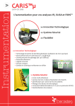
Français Fr. AVRIL. 2013 FIDIS anti-CCP MX 104 DEFINITION Le coffret FIDIS anti-CCP ( ) constitue une méthode d’identification semi-quantitative d'autoanticorps sur supports particulaires utilisant un système de détection par cytométrie de flux. Il permet la recherche simultanée de 4 anticorps, d’isotype IgG, dirigés contre 4 peptides citrullinés : HCP1, HCP2, VCP1 et VCP2. Ces 4 peptides synthétiques sont issus de protéines humaines (HCP1 et HCP2), et virales (VCP1 et VCP2). Les anticorps détectés appartiennent à la famille des anticorps anti-peptides citrullinés (ACPA) et sont capables de détecter les peptides cycliques citrullinés (CCP) ainsi que le fibrinogène déiminé. VALEUR DIAGNOSTIQUE La polyarthrite rhumatoïde (PR) est l’une des maladies auto-immunes systémiques la plus répandue au monde. Sa prévalence atteint 0,5 à 1,0 % de la population adulte. Le diagnostic de la polyarthrite rhumatoïde repose essentiellement sur des caractéristiques cliniques, radiologiques et immunologiques. Le test sérologique le plus commun est la mesure du facteur rhumatoïde (FR) dans le sérum. Bien que le test du FR présente une bonne sensibilité, ce paramètre manque de spécificité. Les FR sont également retrouvés chez des patients atteints d'autres maladies rhumatismales, inflammatoires ou auto-immunes ou encore d'infections chroniques, et chez des personnes saines. En 1998, Schellekens et ses collaborateurs ont signalé que les auto-anticorps réactifs aux peptides synthétiques linéaires contenant de la citrulline étaient très spécifiques de la PR. Les études ultérieures ont démontré que les variantes cycliques de ces peptides linéaires, appelées peptides cycliques citrullinés (CCP) étaient aussi spécifiques de la PR, mais avec une sensibilité supérieure aux peptides linéaires. En 2008, la recherche des anticorps anti-CCP a été introduite à la Nomenclature des Actes de Biologie Médicale en France. En 2010, l’American College of Rheumatology (ACR) et l’European League Against Rheumatism (EULAR) ont publié conjointement de nouveaux critères de classification pour la polyarthrite rhumatoïde. Pour la première fois, la mesure des ACPA (Anti-Citrullinated Peptides Antibodies), tels que les anti-CCP a été intégrée à ces critères. PRINCIPE DU TEST FIDIS anti-CCP ( ) repose sur l’utilisation de microsphères de taille uniforme différemment colorées et d’un cytomètre en flux interfacé avec un système informatique de digitalisation et de traitement du signal. Une diode rouge du cytomètre en flux, en classant chaque catégorie de microsphères sur la base de sa fluorescence unique (du rouge à l’orange), permet l’identification du paramètre analysé. Parallèlement un laser vert excite la fluorescence d’un composé secondaire pour quantifier la réaction spécifique qui y est associée. Chaque antigène, nécessaire à la réalisation du test (peptides synthétiques : HCP1, HCP2, VCP1 et VCP2), est couplé à une catégorie de microsphères colorées. Les différentes catégories sont ensuite mélangées pour constituer le réactif final, support des réactions immunologiques spécifiques avec les différents autoanticorps recherchés. Le coffret FIDIS anti-CCP ( ) permet de détecter 4 anticorps spécifiques : les anticorps antiHCP1, anti-HCP2, anti-VCP1 et anti-VCP2. Le test est réalisé dans une microplaque de filtration de 96 puits. Il a été montré que les anti-CCP peuvent être mis en évidence très tôt au cours de la maladie, souvent en l'absence de symptômes cliniques, et un nombre important de publications indique que des taux élevés d'anticorps anti-CCP peuvent permettre de pronostiquer le développement d'une maladie érosive. Ces résultats suggèrent un rôle important des anti-CCP dans le diagnostic de la PR à un stade précoce de la maladie. Notice technique, FIDISTM anti-CCP, MX 104, Page 1/14 • Au cours d’une première étape, les échantillons dilués des patients à tester sont incubés en présence des microsphères. Si l’échantillon contient un ou plusieurs anticorps recherchés, ceux-ci vont se fixer au(x) antigène(s) correspondant(s) sur les différentes catégories de microsphères • Après incubation, un lavage par filtration permet d'éliminer les éléments non fixés. • Un anticorps secondaire conjugué à la phycoérythrine dirigé contre les immunoglobulines humaines d’isotype IgG permet de révéler les anticorps précédemment capturés. • Une étape finale de lavage stoppe la réaction et permet d’éliminer les anticorps non liés. • La réaction est alors mesurée par le cytomètre en flux qui identifie chaque type de microsphères et mesure la fluorescence moyenne du conjugué fixé. • Un système de calibration permet, par interpolation, de définir la valeur de l’échantillon en unité arbitraire (UA/ml) sur chacune des spécificités antigéniques suivantes : HCP1, HCP2, VCP1, et VCP2. Le coffret FIDIS anti-CCP ( ) peut être utilisé avec l’automate de dilution/répartition CARIS . PRECAUTIONS D'EMPLOI - Les réactifs en solutions contiennent comme agent de conservation, de l’azide de sodium à une concentration <0.1% (w/v) ou du ProClin® 300 à 0.02% (w/v). Ne pas avaler et éviter tout contact avec la peau et les muqueuses. L’azide de sodium peut former des mélanges explosifs lors de son élimination dans les canalisations de cuivre ou de plomb. Rincer abondamment lors de telles éliminations. - Ne pas utiliser les réactifs si des signes de contaminations ou de modifications apparaissent. - Le coffret FIDIS anti-CCP ( ) a été élaboré dans le respect des Directives européennes 67/548/CEE et 1999/45/CE en ce qui concerne la classification, l’emballage et l’étiquetage des préparations dangereuses. - FIDIS anti-CCP ( ) a été optimisé pour les conditions opératoires précisées dans cette notice. Le non-respect des dilutions, de la préparation des réactifs, du protocole ou la substitution d’un réactif par un autre produit peuvent affecter les performances finales du test. - Les contrôles et le calibrateur sont d’origine humaine. Pour chacun, les recherches d’anticorps anti-HIV 1 et 2, anti-HCV et d’antigènes de l’hépatite B se sont révélées négatives. S’agissant de produits potentiellement infectieux, il est toutefois nécessaire de les manipuler avec les précautions d’usage. STABILITE ET CONDITIONS DE CONSERVATION - Tous les réactifs doivent être conservés entre +2°C et +8°C dans leur conditionnement d’origine. Ne pas congeler les réactifs. Ne pas utiliser un coffret dont les dates de péremption sont dépassées. Remettre immédiatement entre +2°C et +8°C, les réactifs non utilisés. COMPOSITION DU COFFRET Plaque de 96 micropuits à membrane filtrante munie d’un couvercle. Flacon (A) de 4 catégories de microsphères colorées sensibilisées par les peptides synthétiques : HCP1, HCP2, VCP1 et VCP2. Lyophilisées (à reconstituer avec le tampon D) 1 plaque qsp 6ml Flacon (B) tampon de dilution des échantillons (flacon blanc) Prêt à l’emploi 2 x 115ml Flacon de calibrateur* Prêt à l’emploi Les titres sont indiqués sur l’étiquette du flacon. 1 x 1,5ml Flacon de contrôle positif donnant une réactivité standardisée et constituant un contrôle de réaction destiné à vérifier l’activité des réactifs et le bon fonctionnement de l’essai. A diluer Les valeurs attendues sont indiquées sur l’étiquette du flacon. 1 x 250µl Flacon de contrôle négatif A diluer 1 x 250µl Flacon de conjugué anti-IgG humaine couplé à la phycoérythrine. Prêt à emploi 1 x 12ml Flacon (C) de tampon de lavage (flacon noir) Prêt à emploi Flacon (D) de tampon de reconstitution des microsphères Prêt à l’emploi 1 x 100ml 1 x 6ml Les titres* du calibrateur sont exprimés en unités arbitraires par ml (UA/ml). Notice technique, FIDISTM anti-CCP, MX 104, Page 2/14 Préparation des réactifs MATERIEL NECESSAIRE NON FOURNI - Pipettes de précision capables de délivrer précisément de 5µl à 1000µl. Pipette multicanaux ou distributeur répétitif capables de délivrer précisément de 40µl à 300µl ou 5µl à 2ml. Agitateur Chronomètre Sonicateur Papier absorbant Pipettes sérologiques Films pour microplaques ÉCHANTILLONS - - - Utiliser du sérum. Eviter d'utiliser des sérums lipémiques ou hémolysés, ainsi que des prélèvements congelés et décongelés plus d'une fois. Si le dosage n'est pas effectué immédiatement, les échantillons devront être conservés réfrigérés entre +2°C et +8°C pendant 5 jours maximum. Au-delà ils devront être congelés à - 20°C. Afin de limiter toute fixation non spécifique, il est conseillé de centrifuger et de filtrer les échantillons congelés depuis plus de 6 mois et troubles. PREPARATION DU TEST TM Préparation de l’unité de filtration FIDIS Vérifier les tuyaux reliant la pompe au support et le réglage du manomètre (molette totalement fermée). Utilisation du système d’analyse FIDIS TM MLX-BOOSTER TM et du logiciel Se reporter au Manuel d’utilisation fourni avec le TM système FIDIS pour effectuer les étapes de mise en route et de calcul. Pour toutes informations complémentaires et/ou résolution de problèmes, veuillez prendre contact distributeur. auprès de ou de votre 1. Commencer la série comme indiquée dans le Manuel d’utilisation. N.B : L’appareil met 30 minutes pour chauffer après avoir été allumé. Un nouveau préchauffage est nécessaire après 4 heures d'inactivité du système. 2. Les étapes de calibration et de contrôles sont décrites dans le Manuel d’utilisation. Ces 2 étapes devront être exécutées régulièrement 1 fois/mois et à chaque nouveau lot de « Sheath fluid » afin d’assurer une performance optimale de l’appareil. L’étape de calibration doit également être effectuée si la température indiquée sur l’écran de la série du lot est en dehors de celle paramétrée. 3. Programmer un lot ou une série de lots multiples sur TM FIDIS comme indiqué dans le Manuel d’utilisation. 4. Placer la microplaque dans le support de plaque du TM système FIDIS comme indiqué dans le Manuel d’utilisation. 5. Effectuer l’analyse des résultats comme indiqué dans le Manuel d’utilisation. Ramener l’ensemble des réactifs à température ambiante (+18°C/+25°C) avant de les préparer extemporanément. 1. Préparation des microsphères Reconstituer la solution de microsphère en ajoutant tout le flacon D de tampon de reconstitution des billes au flacon A. Attendre 5 minutes et vortexer. Durée de conservation après reprise : 2 mois entre +2°C et +8°C. 2. Préparation des échantillons et des contrôles - Diluer les échantillons et les contrôles au 1/201 dans le tampon de dilution (B). Exemple : 10µl d’échantillon dans 2000µl de tampon de dilution (B). - Agiter vigoureusement au vortex. 3. Définition de la configuration du test Utiliser la feuille de travail contenue dans le coffret pour noter la localisation des échantillons. a. Prévoir systématiquement : 1 puits « blanc réactif » 1 puits pour le contrôle négatif 1 puits pour le contrôle positif 2 puits « calibrateur » b. Détermination du nombre exact de puits nécessaires et de leur attribution Les schémas qui suivent donnent 2 exemples de configuration selon le nombre de puits nécessaires et la disponibilité en puits vierges. Le sens du dépôt doit s’effectuer systématiquement par colonne sans intercaler de puits vides. Une même plaque peut être utilisée au cours de différents essais si les puits non utilisés sont protégés par des films pour microplaque. Un même puits ne peut être réutilisé plusieurs fois. Afin d’éviter toute erreur dans ce sens, il est conseillé d’identifier les puits déjà usagés (voir exemple 2 cidessous). c. Programmation du protocole d’analyses au niveau du FIDIS Se reporter au manuel d’utilisation FIDIS « Programmation d’un batch ou d’un Multibatch. ». Exemple 1 : - la plaque est totalement vierge : l’attribution des puits débute en A1. 1 2 3 4 Blanc E4 E12 E20 B Ctrl- E5 E13 E21 C Ctrl+ E6 E14 E22 D Cal E7 E15 E23 E Cal E8 E16 E24 F E1 E9 E17 E25 G E2 E10 E18 H E3 E11 E19 A 5 6 7 8 9 10 11 12 6. A la fin de la dernière utilisation journalière de l’appareil, exécuter les opérations de lavage et de désinfection avant d'arrêter le système d’analyse conformément aux instructions d'arrêt décrites dans le Manuel d’utilisation. Notice technique, FIDISTM anti-CCP, MX 104, Page 3/14 Exemple 2 : - 16 puits ont déjà été utilisés : l’attribution des puits débute en A3. 1 3 4 5 6 7 8 9 10 Blanc E4 E12 E20 E28 E36 E44 E52 B Ctrl- E5 E13 E21 E29 E37 E45 E53 C Ctrl+ E6 E14 E22 E30 E38 E46 D Cal E7 E15 E23 E31 E39 E47 E Cal E8 E16 E24 E32 E40 E48 A 2 F E1 E9 E17 E25 E33 E41 E49 G E2 E10 E18 E26 E34 E42 E50 H E3 E11 E19 E27 E35 E43 E51 11 12 MODE OPERATOIRE 1. Distribution des microsphères - Protéger les puits non utilisés avec des films plastiques (si nécessaire). - Déposer 50µl de microsphères dans chaque puits, après avoir préalablement agité le flacon vigoureusement au vortex pendant 20s. REMARQUE : Dans le cadre de l’utilisation de l’automate CARIS , une sonication des microsphères de 5 minutes est préconisée. Dans le cadre de l’utilisation d’un automate de dilution/répartition, les étapes 1 et 2 doivent être inversées : distribuer le sérum dilué puis les microsphères. 2. Incubation des échantillons - Déposer 100µl de tampon de dilution (B) pour le blanc réactif. - Déposer 100µl de contrôles dilués, de calibrateur prêt à l’emploi et d’échantillons dilués. - Laisser incuber 30 minutes à température ambiante sans agitation en recouvrant la plaque et en évitant de la laisser sous la lumière directe. 3. Lavage 1 Laver la plaque en utilisant l’unité de filtration par 2 cycles successifs en tampon de lavage (C) : a. Retirer le couvercle de la microplaque et la positionner sur l’unité de filtration. (vérifier que le robinet « casse vide » est en position fermée). b. Déclencher la pompe. Dès la disparition totale du liquide, arrêter la pompe et supprimer rapidement le vide en ouvrant le robinet « casse vide ». Refermer le robinet « casse vide ». c. Distribuer 300µl de tampon de lavage (C). d. Déclencher la pompe. Dès la disparition totale du liquide, arrêter la pompe et supprimer rapidement le vide en ouvrant le robinet « casse vide ». Refermer le robinet « casse vide ». e. Distribuer 300µl de tampon de lavage (C). f. Déclencher la pompe. Après aspiration totale du liquide, compter 5 secondes supplémentaires, arrêter la pompe et supprimer rapidement le vide en ouvrant le robinet « casse vide ». Refermer le robinet « casse vide ». g. Retirer la microplaque du laveur et éliminer le tampon résiduel en tapant fortement sa base 10 fois sur du papier absorbant. h. Repositionner la plaque sur le laveur et filtrer à nouveau 5 secondes. Arrêter la pompe et supprimer rapidement le vide en ouvrant le robinet « casse vide ». i. Retirer la microplaque du laveur et éliminer le tampon résiduel en tapant fortement sa base sur un papier absorbant. j. Placer ensuite la microplaque sur une surface totalement sèche avant de distribuer le conjugué. 4. Incubation du conjugué - Déposer 100µl de conjugué - Laisser incuber 30 minutes à température ambiante sans agitation, en recouvrant la plaque et en évitant de la placer sous la lumière directe. Le temps d’incubation débute après que le conjugué ait été ajouté à tous les puits. Si ce temps n’est pas respecté, les résultats pourront être erronés. Remarque : le conjugué est sensible à la lumière ⇒ Refermer le flacon après utilisation. 5. Lavage 2 Laver la plaque en utilisant l’unité de filtration par 1 cycle en tampon de dilution (B) : Utiliser de préférence deux réservoirs à réactifs distincts pour les lavages 1 et 2. Ou bien nettoyer et sécher correctement le réservoir entre les deux lavages afin de ne pas mélanger les deux tampons B et C. a. Retirer le couvercle de la microplaque et la positionner sur l’unité de filtration. (vérifier que le robinet « casse vide » est en position fermée). b. Déclencher la pompe. Dès la disparition totale du liquide, arrêter la pompe et supprimer rapidement le vide par ouverture du robinet « casse vide ». Refermer le robinet « casse vide ». c. Distribuer 100µl de tampon de dilution (B). d. Déclencher la pompe. Dès la disparition totale du liquide, arrêter la pompe et supprimer rapidement le vide en ouvrant le robinet « casse vide ». Refermer le robinet « casse vide ». e. Retirer la microplaque du laveur et éliminer le tampon résiduel en tapant fortement sa base sur un papier absorbant. f. Distribuer 100µl de tampon de dilution (B) et procéder à l’analyse de la microplaque. En cas de lecture différée, la plaque devra être analysée dans l’heure qui suit l’ajout de la solution (B). Durant cette période, la plaque doit être conservée à température ambiante et protégée de la lumière directe. 6. Analyse L’effectuer conformément au manuel d’utilisation FIDIS et MLX BOOSTER . Notice technique, FIDISTM anti-CCP, MX 104, Page 4/14 CRITERES DE VALIDATION DES RESULTATS CALCUL DES RESULTATS Le calibrateur, les contrôles positif et négatif doivent être testés dans chaque série d’essai pour s’assurer que tous les réactifs et procédures ont été exécutés correctement. Afin de valider les résultats, tous les critères énumérés ci-dessous doivent être rencontrés. En cas de non-conformité d’un de ces critères, le test devra être considéré comme non valide et l’analyse devra être refaite. a. La valeur du contrôle positif doit être comprise dans les limites de celles indiquées sur les étiquettes des flacons correspondants. b. La valeur du contrôle négatif doit être inférieure à 25 UA/ml. La solution de microsphères contient des billes témoins permettant de vérifier la présence de sérum dans les puits, ainsi que le bon déroulement du test. Un puits présentant un signal-réponse non conforme sera invalidé par le logiciel MLX BOOSTER™. Les résultats sont automatiquement calculés par le logiciel MLX-BOOSTER™ et peuvent être imprimés pour chaque analyse. LIMITES Les sérums hémolysés, lipémiques, ictériques, présentant des taux élevés en IgG monoclonales, des complexes immuns ou des facteurs rhumatoïdes peuvent entraîner des résultats faussement positifs. Les sérums de patients immunodéprimés donneront un résultat non valide. CONTROLE DE QUALITE Il est conseillé d’utiliser des contrôles internes ou externes pour les différentes spécificités. FIDELITE DU TEST INTERPRETATION DES RESULTATS Déterminer pour chacune des spécificités, la présence ou l’absence d’anticorps, selon le tableau suivant : Unités arbitraires (UA/ml) < 25 UA/ml 25 - 30 UA/ml > 30 UA/ml Interprétation Négatif Limite(*) Positif Intra-essai Inter-essais (30 tests dans le même essai) (2 tests dans 20 essais différents) Spécificité antigénique Valeur moyenne (UA/ml) CV (%) Valeur moyenne (UA/ml) CV (%) HCP1 48 6.5 55 9 HCP2 37 8.5 40 8 VCP1 36 6.5 40 9 VCP2 42 7 41 7 (*) Les résultats limites doivent être contrôlés sur un second prélèvement et interprétés en fonction d’examens complémentaires et du contexte clinique. Chaque laboratoire doit établir et conserver ses propres valeurs de plage (normale) de référence, en fonction de la population de patients et d’autres facteurs locaux. Déterminer le statut final de l’échantillon : a. Un échantillon est considéré comme « positif » lorsqu’au moins un des résultats obtenus pour les spécificités HCP1, HCP2, VCP1 et VCP2 est trouvé « positif ». (En d’autres termes, un résultat est considéré comme « positif » si au moins une des spécificités présentes à un titre en anticorps supérieur à 30 UA/ml.) b. Un échantillon est considéré « négatif » lorsque le résultat de chaque spécificité est inférieur à 25 UA/ml. c. Un échantillon est considéré « limite » lorsqu’au moins une des spécificités présente un titre en anticorps compris entre 25 et 30 UA/ml. Notice technique, FIDISTM anti-CCP, MX 104, Page 5/14 BIBLIOGRAPHIE WO2004087747 (2004-10-14): Citrullinated synthetic peptides and uses thereof, Inv: Migliorini Paola. WO/2011/061720 (2011.05. 26): Viral citrullinated peptides and uses thereof, Inv: PRATESI, Federico; MIGLIORINI, Paola. Rouquette A-M, Desgruelles C, Laroche P. Evaluation of the new multiplexed immunoassay, FIDIS, for simultaneous quantitative determination of antinuclear antibodies and comparison with conventional methods. Hematology Laboratory, Tenon Hospital, 75020 Paris, France Am J Clin Pathol., 2003; 120 (5): 676-81. WO/2012/001103 (2012.01. 05) : Histone citrullinated peptides and uses thereof, Inv: PRATESI, Federico; ALCARO, Maria Claudia; CHELLI, Mario; LOLLI, Francesco; PAOLINI, Ilaria; PAPINI, Anna Maria; ROVERO, Paolo; MIGLIORINI, Paola. Pratesi F., Tommasi C., Anzilotti C., Puxeddu I., Sardano E., Di Colo G., Migliorini P. Antibodies to a new viral citrullinated peptide, VCP2: fine specificity and correlation with anti-cyclic citrullinated peptide (CCP) and anti-VCP1 antibodies. Clin. Exp. Immunol. 2011, 164, 337. Anzilotti C, Merlini G, Pratesi F, Tommasi C, Chimenti D, Migliorini P. Antibodies to viral citrullinated peptide in rheumatoid arthritis. J Rheumatol. 2006 Apr;33(4):647-51. Arnett FC, Edworthy SM, Bloch DA, et al. The American Rheumatism Association 1987 revised criteria for the classification of rheumatoid arthritis. Arthritis Rheum 1988;31(3):315-24. Van Venrooij WJ, Hazes JM, Visser H. Anticitrullinated protein/peptide antibody and its role in the diagnosis and prognosis of early rheumatoid arthritis. Neth J Med 2002;60(10):383-8. Nienhuis RL, Mandema E, Smids C. A new serum factor in patients with rheumatoid arthritis. The antiperinuclear factor. Ann Rheum Dis 1964;23:302-05. Young BJ, Mallya RK, Leslie RD, et al. Anti-keratin antibodies in rheumatoid arthritis. Br Med J 1979;2:97-9. Hoet RM, Boerbooms AM, Arends M, et al. Antiperinuclear factor, a marker autoantibody for rheumatoid arthritis: colocalisation of the perinuclear factor and profilaggrin. Ann Rheum Dis 1991;50:611-8. Sebbag M, Simon M, Vincent C, et al. The antiperinuclear factor and the so-called antikeratin antibodies are the same rheumatoid arthritis-specific autoantibodies. J Clin Invest 1995;95:2672-9 Schellekens GA, de Jong BA, van den Hoogen FH, et al. Citrulline is an essential constituent of antigenic determinants recognized by rheumatoid arthritis-specific autoantibodies. J Clin Invest 1998;101(1):273-81 Vossenaar ER, van Venrooij WJ. Anti-CCP antibodies, a highly specific marker for (early) rheumatoid arthritis. Clin Applied Immunol Rev 2004;4:239-62. Pruijn GJ, Vossenaar ER, Drijfhout JW, et al. Anti-CCP antibody detection facilitates early diagnosis and prognosis of rheumatoid arthritis. Current Rheumatology Reviews 2005;1(1):17. Rantapaa-Dahlqvist S, de Jong BA, Berglin E, et al. Antibodies against cyclic citullinated peptide and IgA rheumatoid factor predict the development of rheumatoid arthritis. Arthirtis Rheum 2003: 48(10):2741-49 Meyer O, Labarre C, Dougados M, et al. Anticitrullinated protein / peptide antibody assays in early Rheumatoid Arthritis for predicting five year radiographic damage. Ann Rheum Dis 2003: 62:120-26 Aletaha D, Neogi T, Silman AJ, et al. Rheumatoid Arthritis Classification Ciriteria. An American College of Rheumatology/European League against Rheumatism Collaborative Initiative. Arthritis Rheum 2010: 62 (9):2569-81 Susan S. Copple, Thomas B. Martins, C. Masterson, E. Joly, Harry R. Hill. Comparison of Three Multiplex Immunoassays for Detection of Antibodies to Extractable Nuclear Antigen Using Clinically Defined Sera. Ann. N.Y Acad. Sci. 1109: 464-472 (2007). Buliard A, Fortenfant F, Ghillani-Dalbin P, Musset L, Oksman F, Olsson N.O. Related Articles, Links [Analysis of nine autoantibodies associated with systemic autoimmune diseases using the Luminex technology. Results of a multicenter study]. Ann Biol Clin (Paris), 2005; Jan-Feb, 63 (1): 51-8. Notice technique, FIDISTM anti-CCP, MX 104, Page 6/14 RECAPITULATIF DU MODE OPERATOIRE QUANTITE A REACTIFS DISTRIBUER CONDITIONS D'INCUBATIONS Déposer 50µl de microsphères dans chaque puits Incubation des échantillons 100µl Tampon de dilution (B) : blanc réactif 100µl Contrôle négatif dilué en tampon de dilution (B) 100µl Contrôle positif dilué en tampon de dilution (B) 100µl Calibrateur prêt à l’emploi (en double) 100µl Echantillons dilués en tampon de dilution (B) Lavage 1 30 min. Température ambiante Laver 2 fois au tampon de lavage (C) (300µl/puits) Incubation du conjugué 30 min Conjugué anti-IgG Prêt à l’emploi 100µl Température ambiante Lavage 2 Laver 1 fois avec du tampon de dilution (B) (100µl/puits) Lecture Ajouter 100µl/puits de tampon de dilution (B) et analyser par insertion de la plaque dans le cytomètre de flux En cas de lecture différée, la plaque devra être analysée dans l’heure qui suit l’ajout de la solution (B). Durant cette période, la plaque doit être conservée à température ambiante et protégée de la lumière directe. LEGENDE DES SYMBOLES Risque Biologique Conserver à Numéro de catalogue Lire le manuel d’utilisation Pour le diagnostic in vitro uniquement Numéro de lot Nombre de tests A utiliser avant Communauté Européenne Microplaque Contrôle négatif Microspheres Diluant échantillon Contrôle positif Conjugué IgG Calibrateur Tampon de lavage Tampon de reconstitution des microsphères Test FIDIS Fabricant 4-6 bld de Beaubourg, CS 90138 CROISSY BEAUBOURG 77435 MARNE LA VALLEE CX2 France Tél : 01 64 62 10 12 Fax : 01 64 62 09 66 E-mail : support@theradiag.com Internet : www.theradiag.com Notice technique, FIDISTM anti-CCP, MX 104, Page 7/14 English Ag. APRIL 2013 FIDIS anti-CCP MX 104 INTENDED USE FIDIS™ anti-CCP ( ) is a semi-quantitative homogeneous fluorescent-based microparticles immunoassay using flow cytometery readings. It is designed for the simultaneous detection of 4 autoantibodies (IgG isotype) directed against 4 synthetic citrullinated peptides : HCP1, HCP2, VCP1 and VCP2. HCP1 and HCP2 are from human protein origin whereas VCP1 and VCP2 are from viral protein origin. These autoantibodies are able to detect cyclic citrullinated peptides (CCP) and deiminated fibrinogen and are associated to anti-citrullinated peptide/protein antibodies (ACPA). SUMMARY AND DIAGNOSTIC VALUES Rheumatoid arthritis is one of the most widespread systemic auto-immune diseases. The prevalence of the disease reaches 0.5 to 1% of adult population.The diagnostic of rheumathoid arthritis is essentially based on clinical, radiological and immunological characteristics. The measurement of Rheumatoid Factor (RF) is widely used. Althought RF test is sensitive, the specificity is low. Indeed, RF are found in patients with rheumatoid diseases, inflammatory or autoimmune diseases, as well as patients subject to chronic infections and in healthy population. In 1998, Schellekens et al. showed that autoantibodies directed against synthetic citrullinated peptides were very specific for rheumatoid arthritis. Further studies shown that auto-antibodies directed against cyclic citrullinated peptides were specific for rheumatoid arthritis and were more sensitive than linear peptide. In 2008, the search for anti-CCP antibodies was introduced to the Nomenclature of the Medical Biology’s acts in France. In 2010, the American College of Rheumatology (ACR) and the European League Against Rheumatism (EULAR), published criteria for the classification of rheumatoid arthritis. For the first time, serological criteria included the measurement of anticitrullinated peptide/protein antibodies (ACPA), such as anti-CCP antibodies for example. ASSAY PRINCIPLE FIDIS™ anti-CCP ( ) is based on the use of distinct uniform size color-coded microspheres and a benchtop flow cytometer interfaced to digital signal processing hardware and software. A red diode laser beam in the flow cytometer classifies each set of microspheres on the basis of its unique fluorescence intensity (red to orange) thus identifying which analyte is being tested. At the same time, a green laser beam illuminates the external second molecule fluorescence to quantify the reaction related to the specific analyte. Each antigen (synthetic peptides : HCP1, HCP2, VCP1 and VCP2) required for the assay is coupled to an individual set of microspheres through its surface functional groups. The different peptides coupled microspheres are mixed together, constituting the final microsphere reagent. The FIDIS™ anti-CCP ( ) allows the detection of 4 autoantibody specificities: anti-HCP1, anti-HCP2, anti-VCP1 and anti-VCP2. It has been shown that anti-CCP antibodies can be detected at the onset of the disease, before any kind of clinical symptoms. Several publications show that a high level of anti-CCP antibodies is correlated to a very active disease. These results demonstrate the importance of the anti-CCP antibodies for the diagnosis of rheumatoid arthritis in the early state of the disease. Product Insert, FIDIS TM anti-CCP, MX 104, Page 8/14 The test is performed in a 96 well microplate with a filtering membrane. • In the first step, the sample is distributed in each well containing the microspheres mixture. If this sample contains one or more of the suspected antibodies, this(ese) antibody(ies) bind to the corresponding antigen(s) on the various sets of microspheres. • After incubation, a wash step using a filtration process removes the unbound antibodies. • A phycoerythrin labelled anti-human IgG conjugate is then added that binds to the previously bound antibodies. • A final wash step allows to stop the reaction. • The reaction is then directly measured by the flow cytometer, which differentiates each set of microspheres according to its fluorescence color while simultaneously measuring the average fluorescence emitted by the conjugate. • A calibration system allows the determination of the titer (AU/mL) of each sample by interpolation for each following antigenic specificity : HCP1, HCP2, VCP1 and VCP2. The FIDIS™ anti-CCP kit could be used with CARIS system (diluting and dispensing device). CAUTION - Reagents in solution contain less of 0.1% (w/v) sodium azide and 0.02% (w/v) of Proclin 300. Do not eat and avoid contact with skin and eyes. Azide can form explosive mixtures in copper or lead piping. Rinse thoroughly after flushing. - Avoid to use reagents if signs of contamination or other visible changes occur. - The FIDIS™ anti-CCP ( ) has been developed according to CE Directives 67/548/EEC and 1999/45/EC regarding classification, packaging and labelling of dangerous preparations. - The FIDIS™ anti-CCP ( ) has been optimized for use as described in this procedure. Do not substitute other manufacturer reagents. Dilution or alteration of these reagents may also alter the performance of the test. Follow thoroughly the test procedure to insure optimal performance. - Calibrators and controls are from human origin. The human sera used in the preparation of these products were tested and found non-reactive for antibodies to HIV-1, HIV-2, anti-HCV and Hepatitis B antigen. Because no test method can offer complete assurance that infectious agents are absent, handle as if capable of transmitting infectious diseases. SPECIMEN COLLECTION AND HANDLING The test is performed on serum. Lipemic sera should be avoided, as well as samples which have been frozen and defrosted more than once. If the test is not run immediately, the samples should be stored at +2°C/+8°C for a maximum of 5 days, for longer storage, frozen undiluted samples at -20°C. To avoid non-specific binding, it is recommended to centrifuge and filter the cloudy samples or samples frozen for more than 6 months. - - - REAGENTS SUPPLIED 96 wells microplate with filtering membrane and lid. 1 plate Vial (A) of color-coded microsphere set of 4 sensitized by HCP1, HCP2, VCP1 and VCP2 synthetic peptides. Lyophilized (to be reconstituted with the buffer named qs 6mL D) Vial (B) of sample dilution buffer (white vial) Ready to use 2 x 115mL Vial of calibrator* Ready to use Each titer is printed on the vial label 1 x 1.5mL Vial of positive control* concentrated. This control has a standard reactivity, which provides evidence of the proper reagents activity and proper assay performance. To be diluted Expected values are printed on the vial label. Vial of negative control concentrate To be diluted 1 x 250 µL 1 x 250µL Vial of anti-human IgG coupled to phycoerythrin Ready to use 1 x 12mL Vial (C) of washing buffer (black vial) Ready to use 1 x 100mL Vial (D) of reconstitution buffer for the microsphere set Ready to use 1 x 6mL Calibrator titers* are expressed in arbitrary units per ml (AU/ml). Product Insert, FIDIS TM anti-CCP, MX 104, Page 9/14 ADDITIONAL MATERIAL – NOT SUPPLIED - - Precision pipettes capable of accurately delivering 5µL to 1000µL Multichannel Pipettes or dispensers capable of accurately delivering 40µL to 300µL or 5µL to 2 mL Vortex mixer Laboratory timer to monitor incubation steps Ultrasonic bath Absorbent towel Serological pipettes Microplate sealing films STABILITY AND STORAGE - Store reagents in their original packaging at +2°C to +8°C. Do not freeze reagents. Do not use kits beyond the expiration date. After use, store all components immediately back at +2°C to +8°C. TEST SET-UP FIDIS Washer Check the tubing connecting the pump to the stand and the manometer setting (wheel completely closed). Using FIDIS Analyzer and MLX-BOOSTER™ Software See the User’s Manual provided with the FIDIS Instrument for detailed instructions on running the equipment and for calculation. For additional information and/or trouble shooting problems, please contact subsidiaries or distributors. 1. Start the run as described in the User’s Manual. NOTE: The FIDIS takes 30 minutes to warm up after being turned on. A new warm up is necessary after 4 hours of system inactivity. 2. Calibration and controls are described in the User’s Manual. Calibration and controls should be routinely performed once per month and each time a new lot of Sheath fluid reagent is used in order to insure optimal instrument performance. Calibration should also be performed when the temperature is out of the determined range shown on the run batch screen. 3. Programming a batch or a multibatch of test protocol on the FIDIS as described in the User’s Manual. 4. Load the microplate into the plate holder of the FIDIS as described in the User’s Manual. 5. Analyze the results according the User’s Manual. 6. When finished for the day, perform the sanitizing and soak operations, prior to turning the Analyzer off according to the shutting down procedure described in the User’s Manual. Reagents Preparation Remove the individual components from storage, allow them to warm up to room temperature (+18°C to +25°C), and mix them well. 1. Microsphere preparation - Reconstitute the microsphere solution by adding the vial D of reconstitution buffer to the vial A of lyophilized microspheres. Wait 5 minutes and vortex. - Stable 2 months at +2°C to +8°C, after reconstitution. 2. Preparation of samples and controls. - Dilute the samples and controls at 1:201 in dilution buffer (B). Ex.: 10µL sample in 2000µL dilution buffer (B). - Vortex vigorously. 3. Assay configuration Use the work-sheet included in the kit to identify the location of the samples. a. When setting up the test, systematically take into account the following well requirements: See examples below 1 “reagent-blank” well 1 well for negative control 1 well for positive control 2 “calibrator” wells b. Calculation of the correct number of wells necessary and their location. In the following examples, 2 different configurations are described, according to the number of wells needed, and the availability of unused wells. The sample dispensing must be systematically carried out in a column. Do not leave empty wells. The same microplate can be used for more than one serie of tests if unused wells are protected by microplate sealing film. Any single well can only be used once. To avoid any error in using a well more than once, identify the used wells with a marker (see example 2 below). c. Programming of the test protocol on the FIDIS Refer to the FIDIS user’s manual: “Creating a batch or a multi-batch”. Example 1: - The microplate is totally blank (no well has yet been used): Use A1 as the 1st well. 1 2 3 4 A Blank S4 S12 S20 B Ctrl- S5 S13 S21 C Ctrl+ S6 S14 S22 D Cal S7 S15 S23 E Cal S8 S16 S24 F S1 S9 S17 S25 G S2 S10 S18 H S3 S11 S19 5 Product Insert, FIDIS 6 TM 7 8 9 10 11 12 anti-CCP, MX 104, Page 10/14 Example 2: 16 wells have already been used: Use A3 as the 1st well. - 1 3 4 5 6 7 8 9 10 Blank S4 S12 S20 S28 S36 S44 S52 B Ctrl- S5 S13 S21 S29 S37 S45 S53 C Ctrl+ S6 S14 S22 S30 S38 S46 D Cal S7 S15 S23 S31 S39 S47 E Cal S8 S16 S24 S32 S40 S48 F S1 S9 S17 S25 S33 S41 S49 G S2 S10 S18 S26 S34 S42 S50 H S3 S11 S19 S27 S35 S43 S51 A 2 11 12 ASSAY PROCEDURE 1. Microspheres dispensing - Before to start the assay and if it is necessary, protect the unused wells with a microplate sealing film. - Vortex the microspheres reagent vigorously for 20s and dispense 50µL in each well to be used. PLEASE NOTE: TM If CARIS is used, the microspheres should be sonicated for 5 minutes. If a dispensing/diluting device is used (like CARIS™), the procedure steps 1 and 2 should be reversed: dispense diluted sera in first and then, dispense microspheres 2. Addition and Incubation of the samples - Dispense 100µL of sample dilution buffer (B) for reagent blank. - Dispense 100µL of prediluted controls, of ready to use calibrator and of prediluted samples. - Cover the plate and incubate 30 minutes at room temperature, away from direct sunlight and without shaking. 3. Wash step 1 Wash the plate 2 times with washing buffer (C) using filtration unit: a. Remove the microplate lid (verify if the vacuum break valve is closed) and place it on the washer. b. Start the pump. Stop it when the whole incubation liquid has been removed in totality and open the vacuum break valve. Close the vacuum break valve. c. Dispense 300µL of washing buffer (C) in all the used wells. d. Start the pump. Stop it when the whole buffer has been removed and open the vacuum break valve. Close the vacuum break valve. e. Dispense 300µL of washing buffer (C) in all the used wells. f. Start the pump. Stop it after 5 additional seconds when all the buffer has been removed and open the vacuum break valve. Close the vacuum break valve. g. Remove the microplate from the washer and remove the residual buffer by tapping vigorously the plate 10 times on an absorbent towel. h. Place again the microplate on the washer and start the pump for 5 seconds. Stop it and quickly open the vacuum break valve. i. Remove the microplate from the washer and remove the residual buffer by blotting vigorously the plate on an absorbent towel. j. Place the plate on a totally dry surface before starting the conjugate incubation. 4. Incubation of the conjugate - Dispense 100µL of ready to use conjugate in each well used. - Cover the plate and incubate 30 minutes at room temperature away from direct sunlight and without shaking. The incubation time starts after the conjugate has been added to all wells. If this timing is not followed, the results might be erroneous. Note : the conjugate is photosensitive. ⇒ After using, close the vial carefully. 5. Wash step 2 Wash the plate using the filtration unit on 1 cycle with dilution buffer (B). Use two different reagent reservoirs for the 2 washing steps. Either clean and dry correctly the reservoir between both wash steps (avoid to mix both B and C buffers). a. Remove the microplate lid (verify that the vacuum break valve is closed) and place it on the washer. b. Start the pump. Stop it when the whole incubation liquid has been removed and open the vacuum break valve. Close the vacuum break valve. c. Dispense 100µL of dilution buffer (B) in all the used wells. d. Start the pump. Stop it when the whole buffer has been removed and open the vacuum break valve. Close the vacuum break valve. e. Remove the microplate from the washer and remove the residual buffer by blotting the plate on an absorbent towel. f. Dispense 100µL of dilution buffer (B) in all the used wells. Place the plate on a totally dry surface at room temperature. The reading must be doing in the hour following adding the solution (B). During this time, keep the plate at room temperature away from direct sunlight. 6. Reading of the test Follow the MLX-BOOSTER manual: Processing batches. Product Insert, FIDIS TM TM and FIDIS™ user’s anti-CCP, MX 104, Page 11/14 VALIDATION CRITERIA OF THE RESULTS CALCUL OF RESULTS Calibrator, negative and positive controls have to be run with every batch of samples to ensure that all reagents and procedures performed properly. In order to validate the results, all the criteria listed below must be met. Otherwise, the test is invalid and must be repeated. a. The positive controls should show a value within the limits printed on the corresponding vial labels. b. The negative controls should be less than 25 AU/mL. The microsphere set contains Internal standard beads allowing to verify the presence of serum in wells, as well as the good respect of the test procedure. Wells presenting a not corresponding fluorescent signal will be no valid with MLX BOOSTER software. The results are automatically calculated by MLXBOOSTER™ software and can be printed for each assay. LIMITATION Hemolytic, lipemic, icteric samples or samples with abnormal concentration of IgG and/or complement levels or samples with rheumatoid factor may confound the results of this assay. The serums from Immunodeficient patients will give a no valid result. QUALITY CONTROL It is recommended to use internally and externally sourced control material for the different specificities. INTERPRETATION OF RESULTS REPRODUCIBILITY Determine for each specificity, the presence or the absence of antibodies, according to the following table: Antigenic Specificity Arbitrary Units (AU/mL) Interpretation < 25 AU/mL 25 - 30 AU/mL > 30 AU/mL Negative Equivocal(*) Positive (*) Borderline results should be controlled on a second sample and the interpretation of the results should be done in the frame of additional testing and taking into account the clinical status of the patient. Within-run Between-run (30 tests in the same run) Mean value CV (%) AU/mL (2 tests in 20 different runs) Mean value CV (%) AU/mL HCP1 48 6.5 55 9 HCP2 37 8.5 40 8 VCP1 36 6.5 40 9 VCP2 42 7 41 7 Each laboratory should establish and maintain its own references (normal) range values, based on the patient population and other local factors. Determine the final status of the sample: a. A sample is considered « positive » when at least one result from HCP1, HCP2, VCP1 and VCP2 antibody specificity is found « positive ». (In other terms, a result is considered “positive” if at least one result from each antibody specificity is higher than 30 AU/mL) b. A sample is considered « negative » when the result from each specificity is less than 25 AU/mL. c. A sample is considered « equivocal » when at least one of the result is comprised between 25 AU/mL and 30 AU/mL. Product Insert, FIDIS TM anti-CCP, MX 104, Page 12/14 BIBLIOGRAPHY WO2004087747 (2004-10-14): Citrullinated synthetic peptides and uses thereof, Inv: Migliorini Paola. WO/2011/061720 (2011.05. 26): Viral citrullinated peptides and uses thereof, Inv: PRATESI, Federico; MIGLIORINI, Paola. WO/2012/001103 (2012.01. 05) : Histone citrullinated peptides and uses thereof, Inv: PRATESI, Federico; ALCARO, Maria Claudia; CHELLI, Mario; LOLLI, Francesco; PAOLINI, Ilaria; PAPINI, Anna Maria; ROVERO, Paolo; MIGLIORINI, Paola. Buliard A, Fortenfant F, Ghillani-Dalbin P, Musset L, Oksman F, Olsson N.O. Related Articles, Links [Analysis of nine autoantibodies associated with systemic autoimmune diseases using the Luminex technology. Results of a multicenter study]. Ann Biol Clin (Paris), 2005; Jan-Feb, 63 (1): 51-8. Rouquette A-M, Desgruelles C, Laroche P. Evaluation of the new multiplexed immunoassay, FIDIS, for simultaneous quantitative determination of antinuclear antibodies and comparison with conventional methods. Hematology Laboratory, Tenon Hospital, 75020 Paris, France Am J Clin Pathol., 2003; 120 (5): 676-81. Pratesi F., Tommasi C., Anzilotti C., Puxeddu I., Sardano E., Di Colo G., Migliorini P. Antibodies to a new viral citrullinated peptide, VCP2: fine specificity and correlation with anti-cyclic citrullinated peptide (CCP) and anti-VCP1 antibodies. Clin. Exp. Immunol. 2011, 164, 337. Anzilotti C, Merlini G, Pratesi F, Tommasi C, Chimenti D, Migliorini P. Antibodies to viral citrullinated peptide in rheumatoid arthritis. J Rheumatol. 2006 Apr;33(4):647-51. Arnett FC, Edworthy SM, Bloch DA, et al. The American Rheumatism Association 1987 revised criteria for the classification of rheumatoid arthritis. Arthritis Rheum 1988;31(3):315-24. Van Venrooij WJ, Hazes JM, Visser H. Anticitrullinated protein/peptide antibody and its role in the diagnosis and prognosis of early rheumatoid arthritis. Neth J Med 2002;60(10):383-8. Nienhuis RL, Mandema E, Smids C. A new serum factor in patients with rheumatoid arthritis. The antiperinuclear factor. Ann Rheum Dis 1964;23:302-05. Young BJ, Mallya RK, Leslie RD, et al. Anti-keratin antibodies in rheumatoid arthritis. Br Med J 1979;2:97-9. Hoet RM, Boerbooms AM, Arends M, et al. Antiperinuclear factor, a marker autoantibody for rheumatoid arthritis: colocalisation of the perinuclear factor and profilaggrin. Ann Rheum Dis 1991;50:611-8. Sebbag M, Simon M, Vincent C, et al. The antiperinuclear factor and the so-called antikeratin antibodies are the same rheumatoid arthritis-specific autoantibodies. J Clin Invest 1995;95:2672-9 Schellekens GA, de Jong BA, van den Hoogen FH, et al. Citrulline is an essential constituent of antigenic determinants recognized by rheumatoid arthritis-specific autoantibodies. J Clin Invest 1998;101(1):273-81 Vossenaar ER, van Venrooij WJ. Anti-CCP antibodies, a highly specific marker for (early) rheumatoid arthritis. Clin Applied Immunol Rev 2004;4:239-62. Pruijn GJ, Vossenaar ER, Drijfhout JW, et al. Anti-CCP antibody detection facilitates early diagnosis and prognosis of rheumatoid arthritis. Current Rheumatology Reviews 2005;1(1):17. Rantapaa-Dahlqvist S, de Jong BA, Berglin E, et al. Antibodies against cyclic citullinated peptide and IgA rheumatoid factor predict the development of rheumatoid arthritis. Arthirtis Rheum 2003: 48(10):2741-49 Meyer O, Labarre C, Dougados M, et al. Anticitrullinated protein / peptide antibody assays in early Rheumatoid Arthritis for predicting five year radiographic damage. Ann Rheum Dis 2003: 62:120-26 Aletaha D, Neogi T, Silman AJ, et al. Rheumatoid Arthritis Classification Ciriteria. An American College of Rheumatology/European League against Rheumatism Collaborative Initiative. Arthritis Rheum 2010: 62 (9):2569-81 Susan S. Copple, Thomas B. Martins, C. Masterson, E. Joly, Harry R. Hill. Comparison of Three Multiplex Immunoassays for Detection of Antibodies to Extractable Nuclear Antigen Using Clinically Defined Sera. Ann. N.Y Acad. Sci. 1109: 464-472 (2007). Product Insert, FIDIS TM anti-CCP, MX 104, Page 13/14 SUMMARY OF THE TEST PROCEDURE VOLUME TO BE DISTRIBUTED INCUBATION REAGENTS CONDITIONS Dispense 50µL of microspheres reagent in each well to be used 100µL Dilution buffer (B): reagent blank 100µL Diluted negative control in dilution buffer (B) 100µL Diluted positive control in dilution buffer (B) 100µL Ready-to-use Calibrator (in duplicate) 100µL Diluted samples in dilution buffer (B) Incubation of the samples Washing 1 30 min. Room temperature Wash twice in washing buffer (C) (300µL/well) Incubation 100µL of the conjugate 30 min Ready to use Anti-IgG conjugate Room temperature Washing 2 Wash 1 time in Dilution buffer (B) (100µL/well) Reading Add 100µL/well of Dilution buffer (B) and proceed immediately with the reading in FIDIS™ Instrument The reading must be doing in the hour following adding the solution (B). During this time, keep the plate at room temperature away from direct sunlight SYMBOLS USED Biological risk Temperature limitation Catalog Number Read instructions for use In Vitro Diagnostic Use Lot Number Number of tests Use by EC Declaration of Conformity Microplate Negative control Microspheres Specimen diluent Positive control IgG Conjugate Calibrator Wash Buffer Microsphere reconstitution buffer FIDIS test Manufacturer 4-6 bld de Beaubourg, CS 90138 CROISSY BEAUBOURG 77435 MARNE LA VALLEE CX2 France Tel : +33 (0)1 64 62 10 12 Fax : +33 (0)1 64 62 09 66 E-mail : support@theradiag.com Internet : www.theradiag.com Product Insert, FIDIS TM anti-CCP, MX 104, Page 14/14