1
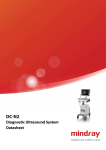
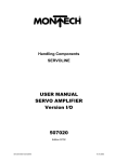
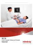
MINDRAY M7 Diagnostic Ultrasound System System Description Transducer Type and Scanning Methods The M7/M7T is an ergonomically designed portable and ease-of-use machine for multi-specialty use like adults, pregnant women, pediatric patients and neonates. It is intended for use in abdominal, gynecology, obstetrics, peripheral vascular, small parts, urological, cardiac, anesthesia, emergency, ICU/CCU, pediatrics and neonates, transcranial Transducer Types Linear Array transducer Phased Array Transducer Convex Array Transducer 4D Volume Transducer Scanning Methods (neonatal Electronic convex Electronic linear Electronic sector cephalic), first aid, interventional, MSK, athletic medical treatment and intraoperative exams. Dimensions and Weight System Introduction Width: 361mm (14.21 inch) Depth: 357mm (14.06 inch) can provide maximum shock protection for system. Height: 75mm (2.95 inch) Weight: approx. 6.5kg (including batteries and 4D M7 is with the total quality Magnesium alloy shell. It System Boot Configuration) board, without power adapter) Electrical Power Monitor Voltage 100-240V~ (AC adapter) Brightness adjustment Screen Saver Open Angle adjustable: 150°(The angle UMT-300 Mobile Trolley) Frequency: 50/60Hz Input power between the monitor and control panel) 1.5- 0.6A (AC adapter) 600 VA (configured with UMT-300 Mobile Trolley) AC adapter output: Voltage: 12V Output current: 10A Battery Lithium-Ion Battery Pack: 11.1V 15 inch LCD, High-Resolution Monitor. Max Resolution:1024 X 768 220-240V~, 50/60Hz (Configured with System Boot Time(From Standby mode):12s (Depend on the Configuration) AC adapter input System Boot Time: 50s (Depend on the , 4500mAh Operating Environment Control Panel Power/Battery Indicator Alphanumeric Keys Function Keys Knobs Ergonomic Soft Key Operation Backlight Keys 8 Segment TGC Blank Keys for User-Defined Functions Trackball: color and Sensitivity Adjustment Ambient temperature: 0°C ~ 40°C Brightness adjustment Relative humidity: 30% ~ 85% (no condensation) Integrated Speakers, Audio Volume Adjustment Atmospheric pressure: 700 hPa ~ 1060 hPa Storage & Transportation Environment Ambient temperature: -20°C ~ 55°C Relative humidity: 30% ~ 95% (no condensation) Atmospheric pressure: 700 hPa ~ 1060 hPa Handle Transducer port 1 port connects to a transducer or the probe extend module. 1/8 Transducer locking lever MINDRAY M7 Diagnostic Ultrasound System Power input port 1 port connect to the power adapter USB Port: 2 I/O(Input/Output) Extend Port Ethernet Port : Connect to the network 1 port connect to the I/O Extend Module S-Video Separate Video Output Probe Extend Module (option) External Wireless Ethernet adapter Support Model: PEM-21 Mobile Trolley: The standard transducer in the main unit extends UMT-200 to three ports by the probe extend module. UMT-300 15-inch Extra LCD Display (Option) The probe extend module need the UMT-300 with Power supply module (Option) the power module when use the 4D probe. I/O Extend Module(iDocTM, option): External DVD R/W Storage (Option) Peripherals Supported Model: IOM-21 M7 support the following peripheral equipments. Apply the additional input and out ports. Including: 2 USB Port 1 ECG Port 1 Serial Port 1 Audio Output Port: Left/Right 1 Mic In port (Connect to the Microphone) 1 Remote Control Port 1 Composite Video Output Port 1 DVI-I (Digital Video Interface-Integrated) Black / White Video Printer (Digital) SONY UP-D897 MITSUBISHI P93DC MITSUBISHI P95DW-N Color Video Printer (Digital): Output Port Should be configured with I/O extend module SONY UP-D23MD Graph / Text Printer HP Deskjet TMD2568 HP OfficeJet TMJ3600 (HP Officejet TMJ3608 All-in-One) V/A Extend Module (option) Model: VAM-21 HP Color LaserJet TMCM1015 Connect to the USB port and apply the additional Mindray only support the following peripheral Video and Audio input ports. Including: equipments as the option. Audio Input Port Left/Right Composite Video Input Port Separate Video Input Port SONY UP-D897 MITSUBISHI P93DC System Application M7 can provide a variety of clinical applications, and ECG module (option) add new applications easily. In every application, user Model: ECG-21 can choose a variety of exam modes. These Connect to the I/O (Input/Output) Extend Port and applications including: apply the ECG function. Abdomen Obstetrics Gynecology ECG Lead port Connection port: to connect to I/O extend Cardiology module Small Parts Urology 2/8 MINDRAY M7 Diagnostic Ultrasound System M –Mode Vascular Pediatrics M-Mode Nerve Free Xros M(Anatomical M ) with Max.3 EM (Emergency) sampling lines Imaging Features Octal beam-former System scanning depth Minimum: 18mm (Probe Dependent) Maximum: 388mm(Probe Dependent ) Color M Mode (CM) Color Doppler Imaging Power Doppler Imaging Power Doppler Imaging Dir-Power Doppler Imaging Spectrum Doppler Gray scale Level:256 PWD (Pulse Wave Doppler) The Max. system frame rate HPRF (High Pulse Repetition Frequency) B mode: ≥640 (Probe Dependent) Color mode: ≥360(Probe Dependent) Display Mode Single Window System processing channels: 2048 Dual-Split System dynamic range: ≥160dB ,A/D 12Bit Quad-Split Maximum frame rate in 4D reaches 30 volumes/s Dual Live System Focusing: Triplex Live Transmission: 8 Steps Reception:Continuous The System Focus Number: 1 – 16 adjustable The System Focus Positions: 16 Max. adjustable The Max. PRF (Pulse Repeat Frequency , Probe dependent) Special Imaging Technique and Functions B Steer for linear transducer. ExFOV Imaging: Extend imaging for convex transducer Trapezoid imaging for linear transducer TDI (Tissue Doppler imaging) Color mode: 14.3kHz TVI (Tissue Velocity Imaging) PW mode: 24kHz TEI (Tissue Energy Imaging) CW mode: 160kHz TVD (TDI-PW) TVM (Tissue Doppler Velocity M Mode) The Max. Flow Velocities (Transducer dependent) Color mode: 226cm/s PW mode: 462cm/s CW mode: 3080cm/s is necessary, Option) Color mode: 7707Hz PW mode: 6600Hz CW mode: 44000Hz of the myocardium Spot: 1-10 Times Pan: 1-10 Times Frame compare Smart 3D (Freehand 3D ) (Option) Rendering Mode: Surface, Mix, Min,X-Ray, Inversion, Magnification factor: TDI Quantitative Analysis Package : Provide the quantitative tools to evaluate the movement state The Max.WF (Wall Filter, 1-7 levels adjustable ) Free Xros CM (Curved Free Xros M Mode) (TDI 3D Navigation: Wire Cage 4D module (Volume transducer is necessary, Option) Rendering Mode: Surface, Mix, Min, X-Ray, Imaging Mode Inversion which are all with wire cage. (4D B-Mode Fundamental Imaging Tissue Harmonic Imaging Phase Shift Harmonic Imaging Navigation) Static 3D (4D module is necessary, Option) Rendering Mode: Surface, Mix, Min, X-Ray, Inversion which are all with wire cage (3D 3/8 MINDRAY M7 Diagnostic Ultrasound System Navigation) TM iScape (Option) Panoramic view imaging function in all type transducers. Maximum iScape TM imaging length: 110cm (Transducer dependent) TM iBeam (Spatial compounding imaging for linear and convex transducers) iClear TM : (Adaptive speckle suppression imaging for all probes) iTouch TM (Instant image optimization technology in B/PW/ CW/Color/TDI mode+ Auto TGC) Power Doppler Imaging Directional Power Doppler Imaging Tissue Harmonic imaging Phase Shift Harmonic Imaging Trapezoid Imaging iBeam™ iTouch™ iStation™ 320G Integrated Hard Disk Multi-language screen display and control panel overlay TM iZoom DVD-RW Intelligent high quality real-time image zoom in Net Storage Function function. Have the capability to direct transfer data through a network cable to PC or another M7 system. No special software dependent. Travelling case zoom in the image area with the image parameters for full screen) Multi-frequency probes for 2D and Doppler imaging mode System Language Color Doppler Imaging TSI (Tissue Specific Imaging ) (Zoom in the image area only for full screen; Software display and keyboard input available: Chinese/English/German/Spanish/French/Italian /Portuguese/Russian/Czech/Polish Options External USB DVD-RW: SE-S224Q I/O extend module: IOM-21 Probe extend module: PEM-21 V/A extend module: VAM-11 ECG module: ECG-21 ECG lead Footswitch: 971-SWNOM (2-pedal) Keyboard input available only: Icelandic/Norwegian/Swedish/Finnish/Turkish/ Danish Control panel overlay available: SP-997-350 (3-pedal) Mobile trolley UMT-200 Weight: 21kg, Width: 470mm, Depth: 657mm, Chinese/German/Spanish/French/Italian/Portugu ese/Russian/Czech/Polish Operation manual available: Chinese/English/German/Spanish/French/Italian Height selective (not available after installed) Platform: 810-870mm Handle position: 890-950mm UMT-300 /Portuguese/Russian Without LCD module (with power module and DVD): System Configuration Standard Configuration High resolution 15 inch LCD Monitor B Mode M Mode PWD (Pulse Wave Doppler) HPRF (High Pulse Repeat Frequency) 4/8 Weight: 43kg, Width: 514mm,Depth: 653mm Height: 855-1015mm With LCD module (with power module and DVD): Weight: 52kg Width: 514mm Depth: MINDRAY M7 Diagnostic Ultrasound System 653mm report) Height: 1550-1830mm Platform height (adjustable): 855-1010mm Pediatric Package (Including related exam mode, comments, measurements, body marks and Pack report) Handbag Dust-Proof Cover mode, comments, measurements, body marks Battery Pack (LI23I001A) and report) Wireless Ethernet adapter Transducers exam mode, comments, measurements, body Needle-guided brackets marks and report) TM iClear CWD (Continuous Wave Doppler) TM Nerve Blocks Package (Including related exam Emergency Medicine Package (Including related DICOM Basic Function Module (Including: task management, DICOM storage, DICOM print, iScape Module DICOM,Storage commitment, DICOM media Free Xros M (Anatomical M) Smart 3D Module DICOM Worklist 4D Module(Including: Static 3D, Volume DICOM MPPS Transducer is necessary) DICOM OB/GYN structured report DICOM vascular structured report DICOM cardiac structured report DICOM Query/Retrieve IMT (Intima-Media Thickness) TDI(Tissue Doppler imaging) storage (including DICOM DIR), etc.) Module (Including: TVI,TEI,TVM) Free Xros CM (Curved Anatomical M-Mode, Display Annotations TDI is necessary) Manufacturer logo TDI Quantitative Analysis(TDI Module is Hospital name necessary) Exam date Abdominal Package (Including related exam Exam time mode, comments, measurements, body marks Acoustic output indices: MI, TIC, TIS, TIB and report) Freeze icon Obstetrical Package (Including related exam Gender mode, comments, measurements, body marks Age and report) ID Gynecological Package (Including related exam Name mode, comments, measurements, body marks Transducer model and report) Current exam mode Cardiac Package (Including related exam mode, ECG icon comments, measurements, body marks and Accession report) Operator Small Parts Package (Including related exam Menu mode, comments, measurements, body marks Image and report) ECG trace Urological Package (Including related exam Transducer orientation mark mode, comments, measurements, body marks Time line and report) Coordinate axis, including depth, time, velocity/ Vascular Package (Including related exam mode, comments, measurements, body marks and 5/8 frequency TGC curve MINDRAY M7 Diagnostic Ultrasound System Focus Vascular Comment Urology Body Mark SMP Measure caliper Nerve Blocks Gray/ Color scale bar EM Thumbnail Cine icon New Trackball functionality status icon Copy Help information Export Soft Menu Load Status icons Delete Biopsy guideline Edit Measure result window Image parameters Storage and Connectivity iStationTM (Intelligent Patient Data Management Unit) Integrated search engine for patient data. Comments and Body Mark Comment Text comment Comment text (Vary by Option), including: Abdomen OB Detailed patient information view Intelligent data backup/restore Patient data/image sending Patient data delete Cardiology Vascular Nerve blocks EM USB ports Image archive on Hard Disk and DVD, temporary saving in cine memory Cine loop Real-time save to hard disk Arrow size adjustable Retrospective Arrow position adjustable Trace Control panel operation (Track ball) Abdomen OB Cardiology GYN 6/8 1-120 cycles, Thumbnail Single image formats: BMP, JPG, DCM, FRM (supports off-line analysis) Multi-frame images formats: AVI, DCM, CIN Mark are related with the application package. Application package (Option) or Prospective (1-120s, or 1-120 cycles, presettable) Mark In M7 system, amount and type of the Body (1-120s, presettable); Arrow orientation adjustable Patient task management External DVD-R/W (Optional) Arrow Body User-Defined Comments Add & Delete Recycle Bin CD-R, CD-RW, DVD-R, DVD-RW) Pediatrics 320G Integrated Hard Disk (DVD+R, DVD+RW, SMP Exam managing: create new exam, activate exam and continue exam GYN Urology User-defined (supports off-line analysis) Storage area: Image area: 640×480 Standard area: 800×600 Full-screen: 1024×768 MINDRAY M7 Diagnostic Ultrasound System iVisionTM Acceleration Cine review: Auto (Auto review segment can be D Trace set), Manual, Simultaneous review for 2D, M/D PS/ED images. Send/ Print image after End Exam Max. frames in HDD ≥10,000,000 frames ≥100,000 frames (JPG format) (FRM format) DICOM: DICOM Storage DICOM print DICOM Worklist Query/ Retrieve Structured Report (SR) Storage Commitment MPPS Media review *Calcification and Study item please refer to the user manual. Diagnostic Report View/add images Edit report Obstetric/vascular analysis Fetal growth curve Print report Import/export report View history report Transducer List C5-2s Convex, Biopsy Guide Available Application: Measurement* Caliper 2D-Mode(B-Mode) Depth Cavity Convex, Biopsy Guide Available Application: Gynecology, Obstetrics, Urology Volume Application: Gynecology, Obstetrics, Urology 6C2s Parallel Micro Convex , Biopsy Guide Available T Length (Trace Length) Application: Ration (D) Cephalic, Nerve, Vascular Pediatric, Cardiac, Abdominal Volume B-Profile Application: Abdominal, OB/GYN VF Diam (Vessel Flow Diameter) L14-6s Linear Biopsy, Guide Available M-Mode Application: HR Slope Small parts, Vascular, Musculoskeletal, Abdominal, Cephalic Pediatric L14-6Ns Distance Linear Biopsy, Guide Available Time Application: Velocity Doppler mode Abdominal, 4CD4s B-Hist (B-Histogram) Color Vel (Color Velocity) 7/8 V10-4Bs Cavity Convex, Biopsy Guide Available Ratio (A) V10-4s Area Cross Obstetrics, Abdominal, Vascular, Nerve Distance Angle Gynecology, Small parts, Vascular, Musculoskeletal, Abdominal, Cephalic Pediatric L12-4s D Velocity Linear, Biopsy Guide Available HR Application: Small parts, Vascular, Orthopedics, Time Musculoskeletal, Abdominal, Cephalic, Pediatric MINDRAY M7 Diagnostic Ultrasound System marking (0123) is the number of the EU-notified body that 7L4s certified meeting the requirements of Annex II of the Linear, Biopsy Guide Available Directive. Application: Small parts, Vascular, Orthopedics, Not all features or specifications described in this document Musculoskeletal, Abdominal, Cephalic, Pediatric may be available in all Transducers and/or modes. L7-3s No part of this manual may be copied or printed, in whole or in part, without written permission. Linear, Biopsy Guide Available The contents of this manual are subject to change without Application: Small parts, Vascular, Abdominal, prior notice and without our legal obligation. Pediatric Note: the contents in this datasheet are applied to Version 2.0 of system software for M7/M7T diagnostic ultrasound 7L5s system. Linear, Biopsy Guide Available Application: Small organ, Vascular Orthopedics, Nerve, Musculoskeletal, P4-2s Phase Array, Biopsy Guide Available Application: Cardiac, Abdominal, Pediatric, Trans cranial P7-3s Phase Array Application: Cardiac, Abdominal, Pediatric, Cardiac, Abdominal, Pediatric, Transcranial P12-4s Phase Array Application: Transcranial CW2s Pencil probe Application: Transcranial, cardiology, pediatrics Safety & Conformance Quality Standards ISO 9001:2000 ISO 13485:2003 Design Standards UL 60601-1 CSA C22.2 No. 601-1 EN 60601-1 and IEC 60601-1 EN 60601-1-1 and IEC 60601-1-1 EN 60601-1-2 and IEC 60601-1-2 EN 60601-2-37 and IEC60601-2-37 EN60601-1-4 and IEC60601-1-4 EN60601-1-6 and IEC60601-1-6 CE Declaration M7/ M7T system is fully in conformance with the Council Directive 93/42/EEC Concerning Medical Devices, as amended by 2007/47/EC. The number adjacent to the CE 8/8