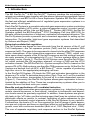
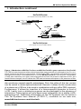
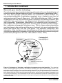
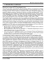
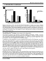
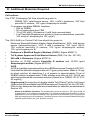
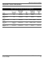
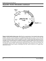
1
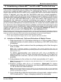
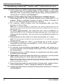
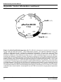
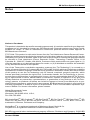
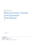
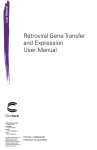
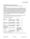
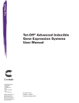
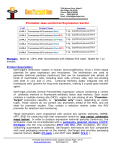
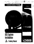
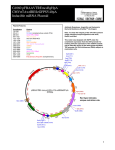
BD RevTet™ System
User Manual
Cat. No. 631020 or K1626-1
631021 or K1627-1
PT3223-1 (PR33666)
Published 27 March 2003
BD Biosciences
BD RevTetTM System User Manual
Table of Contents
I. Introduction
4
II. List of Components
10
III. Additional Materials Required
11
IV. RevTet Procedural Overview
13
V. Plasmid Manipulations
14
VI. Guidelines for Working with Retroviruses
15
VII. Cell Culture Guidelines
16
VIII. Pilot Experiments
18
IX. Virus Production
21
X. Establishing a Stable BD Tet-OffTM or BD Tet-OnTM Cell Line
23
XI. Establishing a Double-Stable, Inducible Cell Line
26
XII. References
29
XIII. Related Products
30
Appendix: Vector Information
31
Note: The viral supernatants produced by these retroviral systems could, depending on your DNA insert, contain potentially hazardous recombinant virus.
Due caution must be exercised in the production and handling of recombinant
retrovirus. The user is strongly advised not to create retroviruses capable of
expressing known oncogenes in amphotropic or polytropic host range
viruses.
Appropriate NIH, regional, and institutional guidelines apply, as well as guidelines
specific to other countries. In the U.S., NIH guidelines require that retroviral
production and transduction be performed in a Biosafety Level 2 facility. For more
information, see appropriate HHS publications. Section VI in this User Manual
contains a brief description of Biosafety Level 2 as well as other general
information and precautions. For more complete information refer to Biosafety in
Microbiological and Biomedical Laboratories available on the world wide web at:
http://bmbl.od.nih.gov
BD Biosciences Clontech
2
www.bdbiosciences.com
Protocol # PT3223-1
Version # PR33666
BD RevTetTM System User Manual
Table of Contents continued
List of Figures
Figure 1.
Mechanism of RevTet-On and RevTet-Off gene expression
5
Figure 2.
Packaging of infectious, replication-incompetent
retroviral particles
6
Figure 3.
Figure 4.
Establishing an inducible cell line with the RevTet System
Infection of 3T3 cells with pRevTet-Off-IN produces a higher
percentage of highly inducible clones
8
9
Figure 5.
Overview of the RevTet procedure
13
Figure 6.
Dose-response curve for the CHO-AA8-Luc Control Cell Line
19
Figure 7.
Fold induction of luciferase activity in different lots of FBS
19
Figure 8.
pRevTet-On Vector map
32
Figure 9.
pRevTet-Off Vector map
33
Figure 10. pRevTet-Off-IN Vector map
34
Figure 11. pRevTRE Vector map and MCS
35
Protocol # PT3223-1
Version # PR33666
www.bdbiosciences.com
BD Biosciences Clontech
3
BD RevTetTM System User Manual
I. Introduction
The BD RevTet-OnTM & BD RevTet-OffTM Systems combine the advantages of
retrovirus-mediated gene transfer with the proven tetracycline-regulated control
of BD Tet-OnTM and BD Tet-OffTM Gene Expression Systems. BD RevTetTM allows
the fast and efficient establishment of regulated gene expression systems in a
wide variety of cell types.
Each RevTet System is a complete retroviral gene expression system containing
a retroviral regulator (r)tTA vector, a retroviral response vector, a control vector,
and a packaging cell line. Like our BD Retro-XTM System (#631508), the RevTet
Systems contain the BD RetroPackTM PT67 Packaging Cell Line (#631510) for
the safe, efficient production of infectious, replication-incompetent retrovirus. The
RevTet Systems provide a powerful and convenient approach to setting up the
tetracycline (Tc)-inducible, high-level gene expression systems first described
by Gossen and Bujard (1992).
Retroviral-mediated tet-regulation
The Tet Systems are based on two elements from the tet operon of the E. coli
Tn10 transposon—the Tet repressor protein (TetR) and the tet operator DNA
sequence (tetO). The gene to be expressed is cloned into the pRevTRE response
vector downstream of the tetracycline-responsive element (TRE), which contains seven direct repeats of the 42-bp tetO sequence and the minimal immediate early promoter of cytomegalovirus (PminCMV). The two systems differ in their
regulatory vector (Figure 1). The RevTet-Off System uses the pRevTet-Off Vector, which contains the tTA regulatory element, a fusion of TetR and the VP16
activation domain. The BD RevTet-OnTM System uses pRevTet-On, based on the
reverse tTA (rtTA). Both systems use the pRevTRE response vector. All the RevTet
Vectors are derived from pLNCX, a BD Retro-XTM vector capable of producing
high-titer virus in the appropriate packaging cell lines.
In the RevTet-Off System, tTA binds the TRE and activates transcription in the
absence of Tc or the Tc derivative doxycycline (Dox). Thus, as Tc or Dox is added
to the culture medium, transcription is turned off in a dose-dependent manner
(Figure 1). Conversely, the RevTet-On System allows gene expression to be activated by the addition of Dox. These two complementary systems allow you to
choose the one that best suits your needs.
Benefits and applications of Tc-mediated induction
In most inducible mammalian gene expression systems (e.g., induction by heavy
metals, steroid hormones, or heat shock), induction is nonspecific and expression levels cannot be precisely regulated. In addition, these systems are generally leaky in the “off” state, and the inducing agent itself may be cytotoxic or have
pleiotropic effects on results. In contrast, regulation of gene expression by the
heterologous bacterial control elements in the RevTet Systems is very specific.
Furthermore, the levels of Tc or Dox required for the full range of gene expression are not cytotoxic and have no significant effect on cell proliferation or animal
growth, even with continuous treatment (Bohl et al., 1997; Mayford et al., 1996).
The use of separate regulatory and response vectors in the RevTet Systems
BD Biosciences Clontech
4
www.bdbiosciences.com
Protocol # PT3223-1
Version # PR33666
BD RevTetTM System User Manual
I. Introduction continued
RevTet-Off System
(Transcription turned off by addition of Tc or Dox)
PCMV
tetR
VP16
copy of pRevTet-Off
regulatory plasmid
VP16
TetR
+ Tc (or Dox)
– Tc (or Dox)
tTA
VP16
Transcription
TetR
PminCMV
TRE
Gene of interest
copy of pRevTRE response plasmid
RevTet-On System
(Transcription turned on by addition of Dox)
PCMV
rtetR
VP16
copy of pRevTet-On
regulatory plasmid
VP16
rTetR
– Dox
+ Dox
rtTA
VP16
Transcription
rTetR
TRE
PminCMV
Gene of interest
copy of pRevTRE response plasmid
Figure 1. Mechanism of BD RevTet-OnTM and BD RevTet-OffTM gene expression. RevTet-Off:
The TRE is located upstream of the minimal immediate early promoter of cytomegalovirus (PminCMV),
which is silent in the absence of activation. tTA binds the TRE—and thereby activates transcription
of the gene of interest—in the absence of Tc or Dox. BD RevTet-OnTMB: The “reverse” Tet repressor
(rTetR) was created by four amino acid changes that reverse the protein’s response to Dox. As a
result of these changes, the rtTA binds the TRE and activates transcription in the presence of Dox.
also produces several specific advantages. First, the ratios of the two vectors
can be varied to obtain optimal induction. Second, it allows for the construction
of a reference (r)tTA line to be used in combination with any pRevTRE construct.
Furthermore, it allows for insertion of a tissue-specific promoter in front of
(r)tTA. Lastly, toxic or deleterious genes can be packaged in the absence of
expression. Transcription will only begin upon integration into a stable (r)tTAexpressing line. Additional information on the Tet Systems can be found in
The Tet Systems User Manual (PT3001-1), available on our web site,
www.bdbiosciences.com/clontech.
Protocol # PT3223-1
Version # PR33666
www.bdbiosciences.com
BD Biosciences Clontech
5
BD RevTetTM System User Manual
I. Introduction continued
Retroviral gene transfer technology
Current retroviral gene transfer technology is based on the coordinated design of
retroviral vectors and packaging cell lines. The development of packaging lines—
cell lines that package retroviral RNAs into infectious particles without the
concomitant production of replication-competent virus—created a new level of
safety and control (Figure 2; Mann et al., 1983; Miller & Buttimore, 1986). To make
a packaging cell line, the structural genes necessary for particle formation and
replication, gag, pol, and env, are integrated into the genome without any other
viral sequences. Subsequent introduction of a retroviral vector containing Ψ+,
transcription and processing elements, and the gene of interest produces hightiter, replication-incompetent infectious virus. That is, these retroviral particles
can infect target cells and transmit the gene of interest, but cannot replicate within
these cells since they lack the viral genes. The separate introduction and
integration of the viral genes into the packaging cell line minimizes the chances
of producing replication-competent virus due to recombination events during cell
proliferation (Morgenstern & Land, 1990; Miller & Chen, 1996).
Packaging Cell
(produces viral proteins from
stably integrated genes)
1) Transfection
2) Integration
Retroviral
vector
Ψ+ Gene X Neor
transient
expression
DNA
Ψ+ Gene X Neor
3) Transcription
RNA
viral
proteins
4) Viral proteins
recognize Ψ+
5) Packaging
6) Budding of infectious but
replication-incompetent virus
Figure 2. Packaging of infectious, replication-incompetent retroviral particles. The retroviral
vector, containing the gene of interest, an antibiotic selection gene (Neor in this example), and Ψ+, the
packaging signal necessary for retrovirus particle formation, can stably integrate or be transiently
expressed. The packaging cell line provides the genes necessary for particle formation which have
been deleted from the vector: gag (core structural proteins), pol (reverse transcriptase, integrase), and
env (coat glycoproteins). Virus released from this cell line contains the products of these genes (and
is infectious), but lacks the genes themselves, thus preventing retroviral production from subsequently
infected cell lines.
BD Biosciences Clontech
6
www.bdbiosciences.com
Protocol # PT3223-1
Version # PR33666
BD RevTetTM System User Manual
I. Introduction continued
Broad host range of PT67-packaged virus
The viral env gene, expressed by the packaging cell line, encodes the envelope
protein which determines the cellular host range of the packaged virus. The
envelope protein allows infection of different cell types through recognition of
specific cellular receptors. The RevTet System includes the RetroPackTM PT67
Packaging Cell Line, an NIH/3T3-based line expressing the 10A1 viral envelope.
Virus packaged from PT67 cells can enter cells via two different surface molecules, the RAM1 and GALV receptors, and thus exhibits a broader host range
than virus packaged by other lines (Miller & Miller, 1994; Miller, 1996).
The RevTet Systems produce retrovirus capable of infecting nearly all dividing
mammalian cells, including primary cell lines. Cell lines that are typically more
difficult to transfect, such as Jurkat and PC12, have been successfully infected
with RevTet viruses. Please note that retroviral transduction (specifically, nuclear
entry of the viral pre-initiation complex) can only occur in actively dividing cells.
In addition to PT67 cells, you may also use other packaging cell lines offered by
BD Biosciences Clontech to provide different host ranges:
• BD EcoPackTM-293 Cell Line (#C3200-1): Generate ecotropic virus to infect a
broad range of mouse and rat cells.
• BD AmphoPackTM-293 Cell Line (#C3201-1): Generate amphotropic virus to
infect a broad range of mammalian cells.
• GP2-293 Cell Line: Available as part of our Pantropic Retroviral Expression
System (#K1063-1), GP2-293 cells produce virus that can infect the broadest
possible range of mammalian and nonmammalian cells.
Constructing a stable expression cell line using PT67 packaging cells
To create a stable cell line that expresses your gene of interest upon induction,
you perform a series of transfections and infections as diagrammed in Figure 3.
The first step is to make two different retroviruses—one to deliver the pRevTetOff or pRevTet-On regulator construct to the target cells and one to deliver the
pRevTRE response construct, containing your gene of interest (Gene X). To
produce the former virus, pRevTet-Off/On Vector is transfected into the packaging cell line. In parallel, you can package pRevTRE-Gene X by performing a
second transfection. Although you can use virus that is transiently produced by
these cell lines for subsequent steps, we recommend that you use antibiotic
selection to create stable virus-producing cell populations. Viral titers of stable
clones are not dependent on the efficiency of transfection, and can be frozen and
used for subsequent experiments.
When packaging is complete, supernatant containing pRevTet-Off/On virus is
used to infect the target cells. Neomycin selection of this culture yields a
population of cells that have integrated (r)tTA. Individual clones are then isolated
from this population to establish a Tet-Off/On cell line. (If desired, the heterogeneous population can be used instead; however, expression systems with the
lowest background and highest inducibility are obtained after isolating individual
Protocol # PT3223-1
Version # PR33666
www.bdbiosciences.com
BD Biosciences Clontech
7
BD RevTetTM System User Manual
I. Introduction continued
A
Packaging Cells
Target Cells
Transfect packaging cell
line with regulatory and
expression vectors.
Infect target
cells with
collected virus
Neor
pRevTetOff/On
Collect viruscontaining
supernatant
Hygr
pRevTRE
Gene of
interest
Select (Neo)
and screen
Tet-On or Tet-Off cell line
(Premade cell lines are also
available from CLONTECH)
Select (Hyg)
and screen
Double-stable cell line expressing the
target gene under doxycycline control
Figure 3. Establishing a inducible cell line with the BD RevTetTM System. The pRevTet-On or
pRevTet-Off Vector and the pRevTRE response vector (expressing your gene of interest or a library)
are separately transfected into the RetroPack PT67 packaging cell line. The resulting virus-containing
supernatants are used to serially infect target cells—first with pRevTet-Off/On virus to produce a stable
Tet-Off/On cell line, then with pRevTRE virus to integrate your gene of interest. Alternatively, the
supernatants can be used simultaneously to infect target cells (not shown). Expression is induced by
the addition (RevTet-On) or withdrawal (RevTet-Off) of doxycycline or tetracycline. Prior to target cell
infection, high-titer virus producing lines can be established with antibiotic selection.
clones.) We recommend storing frozen stocks of this cell line or population to
express future genes of interest from pRevTRE-Gene X or pTRE2 constructs.
Next, the Tet-Off/On cell line is infected with pRevTRE virus. Hygromycin
selection of this culture yields a population of cells containing all the components
needed for Tet-regulated expression. Single clones with the desired induction
characteristics can be isolated at this point.
As an alternative to producing Tet-regulated cell lines via serial infection with
pRevTet-Off/On and pRevTRE virus, you may infect with both viruses simultaneously. However, this method is only recommended for pilot experiments and for
cell lines such as primary cells that cannot be propagated in culture for an
extended period of time.
BD Biosciences Clontech
8
www.bdbiosciences.com
Protocol # PT3223-1
Version # PR33666
BD RevTetTM System User Manual
I. Introduction continued
A
B
70
80
pRevTet-Off-IN (NIH/3T3)
pRevTet-Off-IN (Swiss 3T3)
70
pTet-Off (NIH/3T3)
60
pRevTet-Off (NIH/3T3)
60
50
% of clones
% of clones
50
40
30
20
40
30
20
10
10
0
0
0–20 fold
21–50 fold
>50 fold
Luciferase induction (–Dox RLU/+Dox RLU)
0–20 fold
21–50 fold
>50 fold
Luciferase induction (–Dox RLU/+Dox RLU)
Figure 4. Infection of 3T3 cells with pRevTet-Off-IN produces a higher percentage of highly
inducible clones. Panel A. Infection of NIH/3T3 or Swiss 3T3 cells with pRevTet-Off-IN. Clones
were selected with 1 mg/ml G418. Panel B. Transfection of NIH/3T3 cells with pTet-Off (from our
non-retroviral-based Tet-Off System) or infection with pRevTet-Off. In both panels, clones generated by plasmid transfection or retroviral infection were analyzed for their ability to exhibit inducible
expression of luciferase from the pTRE-Luc vector, and grouped according to fold-induction.
RLU = relative light units.
TM
The BD RevTet Vectors (see maps in the Appendix)
The RevTet Vectors are based on pLNCX (Miller & Rosman, 1989), a vector
derived from Moloney Murine Leukemia Virus (MoMuLV). These vectors contain
the extended retroviral packaging signal, Ψ+, which promotes high-titer virus
production. See the Appendix for detailed maps.
In addition to the regulatory vectors supplied with the RevTet Systems, we offer
pRevTet-Off-IN (#6136-1) which contains an internal ribosomal entry site (IRES)
located between the regulatory sequence and the neomycin-resistance gene.
The inclusion of the IRES, which allows the simultaneous expression of the tet
regulatory protein with Neor from a single transcript, ensures that all of your
G418-resistant clones also express the regulatory protein. Figure 4 shows that
pRevTet-Off-IN yields a higher percentage of clones capable of high induction in
NIH/3T3 cells (Cunningham et al., 1998). pRevTet-Off-IN is sold separately and
can be used in place of pRevTet-Off in the RevTet-Off System.
Tet-Inducible Retroviral Libraries
BD Biosciences Clontech offers Tet-Inducible Retroviral Libraries that allow you
to efficiently screen cDNA libraries directly in human cells. Furthermore, because
the system is inducible and the inserts are not expressed in the packaging cell
line, you can recover toxic or apoptosis-related genes. See Related Products
(Section XIII) for a list of currently available libraries.
Protocol # PT3223-1
Version # PR33666
www.bdbiosciences.com
BD Biosciences Clontech
9
BD RevTetTM System User Manual
II. List of Components
Store cell lines in liquid nitrogen (–196°C).
Store all plasmids at –20°C.
Store Tet System Approved Fetal Bovine Serum at –20°C.
BD RevTet-OnTM System (#631021)
•
10 µg
pRevTet-On Vector (0.5 µg/µl)
•
10 µg
pRevTRE Vector (0.5 µg/µl)
•
10 µg
pRevTRE-Luc Vector (0.5 µg/µl)
•
1 ml
RetroPack PT67 Cell Line (2 x 10⁶/ml)
•
50 ml
Tet System Approved Fetal Bovine Serum
BD RevTet-OffTM System (#631020)
•
10 µg
pRevTet-Off Vector (0.5 µg/µl)
•
10 µg
pRevTRE Vector (0.5 µg/µl)
•
10 µg
pRevTRE-Luc Vector (0.5 µg/µl)
•
1 ml
RetroPack PT67 Cell Line (2 x 10⁶/ml)
•
50 ml
Tet System Approved Fetal Bovine Serum
The following kit components are also available separately:
•
•
•
•
•
pRevTet-On Vector
pRevTet-Off Vector
pRevTRE Vector
RetroPack PT67 Cell Line
Tet System Approved Fetal Bovine Serum
BD Biosciences Clontech
10
#631007
#631003
#631002
#631510
#631101
www.bdbiosciences.com
Protocol # PT3223-1
Version # PR33666
BD RevTetTM System User Manual
III. Additional Materials Required
Cell culture:
The PT67 Packaging Cell Line should be grown in:
DMEM, 90%; fetal bovine serum, 10%; 4 mM L-glutamine; 100 U/ml
penicillin G sodium; 100 µg/ml streptomycin sulfate.
To prepare complete DMEM, combine:
– 500 ml DMEM
– 50 ml tetracycline-free FBS
– 10 ml 200 mM L-Glutamine (4 mM final concentration)
– 5 ml Penicillin/Streptomycin solution (final concentrations: penicillin
100 units/ml; streptomycin 100 µg/ml)
The CHO-AA8-Luc Control Cell Line should be grown in:
Minimum Essential Medium Eagle (Alpha Modification), 90%; fetal bovine
serum (tetracycline-free), 10%; 2 mM L-glutamine; 100 µg/ml G418;
100 units/ml penicillin G sodium; 100 µg/ml streptomycin sulfate;
100 µg/ml hygromycin B
•
•
•
•
•
•
Dulbecco’s Modified Eagle’s Medium (DMEM; Sigma #D5671).
Tet System Approved Fetal Bovine Serum (FBS; Cat. No. 631101)
200 mM L-Glutamine (Sigma #G7513)
Solution of 10,000 units/ml Penicillin G sodium and 10,000 µg/ml
Streptomycin sulfate (Sigma #P0781)
G418
G418 is available in powdered form from BD Biosciences Clontech (631307).
Note that the effective weight is about 0.7 g per g of powder. Make a 10 mg/
ml stock solution by dissolving 1 g of powder in approximately 70 ml of
DMEM (without supplements). Filter sterilize and store at 4°C. (G418 can
also be purchased as a premade solution [Geneticin®; GIBCO 10131-019,
50 mg/ml]).
Hygromycin (for selection of double-stable Tet-Off and Tet-On Cell Lines)
Hygromycin B is available from BD Biosciences Clontech (Cat. No.631309).
Prior to use, determine the optimal concentration for selection as described in
Section VIII.
Notes on antibiotic selection: For maintenance of most cell lines, 100 µg/ml of G418
or hygromycin is a suitable concentration. For selecting stable transformants in HeLa
and NIH/3T3 cells, we have found 400–500 µg/ml G418 (sometimes up to 1 mg/ml) and
200 µg/ml hygromycin to be optimal. For either purpose, we recommend determining the
optimal concentration for your specific cell line.
Protocol # PT3223-1
Version # PR33666
www.bdbiosciences.com
BD Biosciences Clontech
11
BD RevTetTM System User Manual
III. Additional Materials Required continued
•
Trypsin-EDTA (Trypsin; Sigma T3924)
•
Dulbecco’s phosphate buffered saline (DPBS; Sigma D8662)
•
Cell Freezing Medium (Sigma C6164) with or without Dimethyl
sulfoxide (DMSO; Sigma D2650)
•
Tissue culture plates and flasks
•
Cloning cylinders (PGC Scientific 62-6150-40, -45)
or Cloning discs (PGC Scientific 62-6151-14, -12)
•
µm filters
0.45-µ
Use cellulose acetate or polysulfonic (low protein binding) filters. Do not
use nitrocellulose filters because they bind proteins present in the retroviral
membrane, destroying the virus.
For transient and stable transfections
The transient and stable transfections in this protocol can be performed by
various methods. Reagents will depend on which transfection method you use.
We generally use electroporation or the calcium phosphate method for both
transient and stable transfections. Protocols for other methods can be found in
Freshney (1993) and Ausubel et al. (1994).
For infection of target cells
•
Polybrene (Hexadimethrine Bromide; Sigma H9268)
Inducing Agent
•
Doxycycline (Dox; Cat. No. 631311): We recommend using Dox for inducing
gene expression in the RevTet Systems. In the RevTet-On system, Dox has
a 100-fold higher affinity for rtTA than Tc (Gossen & Bujard, 1995). As a result,
Dox produces much greater activation of the rtTA protein and a higher level of
induction of the target gene.
In contrast, both drugs completely inactivate the tTA regulatory protein in the
BD RevTet-Off System. At BD Biosciences Clontech, we use Dox for all
RevTet System experiments. A significantly lower concentration of Dox is
required for complete activation or inactivation (10 ng–1 µg/ml Dox vs.
1–2 µg/ml Tc).
Subtoxic levels of Tc and Dox:The working concentrations of Tc and Dox
used in the RevTet systems are far below the levels known to have cytotoxic
effects on mammalian cells in culture or in transgenic animals.
BD Biosciences Clontech
12
www.bdbiosciences.com
Protocol # PT3223-1
Version # PR33666
BD RevTetTM System User Manual
IV. RevTet Procedural Overview
Plasmid Manipulations
8–10
days
Cell Line Manipulations
(Section V)
(Section VII)
Transform all plasmids
into E. coli.
Establish cultures of
PT67 packaging cells.
Large-scale plasmid preps.
Subclone gene of interest
into pRevTRE.
Prepare frozen cultures
of PT67 cells.
Pilot Experiments (Section VIII)
• Use CHO-AA8-Luc Tet-Off
Cell Line to determine effective
concs. of Tc or Dox stocks.
• Use CHO-AA8-Luc cells to test
serum for Tc contamination.
2–3
weeks
(Alternatively use Tet-Approved
FBS, #8630-1)
• Titrate G418 and hygromycin
to determine optimal conc. for
selection of your target cells.
• Use pRevTRE-Luc to test target
cells with the RevTet system
by establishing an inducible cell
line expressing luciferase.
Virus Production (Section IX)
4
weeks
RevTet-Off/On Virus
• Produce
• Select
• Titer
• Make frozen cultures
RevTRE-Gene X Virus
• Produce
• Select
• Titer
• Make frozen cultures
Control Viruses (as necessary)
• Produce
• Select
• Titer
• Make frozen cultures
Optional: use transiently produced virus to infect target cells.
3–5
weeks
Target Cell
Infections
(Sections X–XI)
RevTet-Off/On Virus
Create Tet cell line:
• Infect
• Select
• Isolate inducible clones
• Prepare frozen cultures
Optional: Use mixed population
RevTRE-Gene X Virus
Incorporate gene of interest
• Infect
• Select
• Isolate inducible clones
• Prepare frozen cultures
• Proceed with experiments
Optional: Instead of sequential infection, you may simultaneously
coinfect and perform double selection (for instance, when using
primary cell cultures).
Figure 5. Overview of the RevTet Procedure. Be sure to read entire protocol before starting.
Protocol # PT3223-1
Version # PR33666
www.bdbiosciences.com
BD Biosciences Clontech
13
BD RevTetTM System User Manual
V. Plasmid Manipulations
A. Propagation of Plasmids
1. Transform each of the plasmids provided in this kit into a suitable E. coli
host strain (e.g., DH5α) to ensure that you have a renewable source of
DNA.
2. You will need to perform large-scale plasmid preparations of any
plasmid that will be introduced into mammalian cells. To ensure the
purity of the DNA, isolate all plasmids for transfection using a
NucleoBond® Plasmid Maxi Kit (Cat. No. 635933, Cat. No. 635934,
Cat. No.635935) or by banding on a CsCl gradient (Sambrook et al.,
1989).
B. Generating Your Gene-Specific Expression Vector
Generate your pRevTRE-Gene X construct using standard molecular
biology techniques, as described below. For more detailed information, see
Sambrook et al. (1989).
1. Purify the target Gene X fragment by any standard method. The cDNA
or gene fragment must contain an ATG initiation codon. In some cases,
addition of a Kozak consensus ribosome binding site (Kozak, 1987)
may improve expression levels; however, many genes have been
efficiently expressed in mammalian systems without the addition of a
Kozak sequence. The cDNA should not contain a polyadenylation
signal. The inclusion of such sequences between retroviral LTRs can
cause premature polyadenylation during virus transcription which
interferes with the production of vector-containing virions (Coffin &
Varmus, 1996). The fragment can be generated using compatible
restriction sites that are present on either side of the gene and in the
cloning vector. If no such sites are present, the gene fragment can be
generated by PCR with suitable restriction sites incorporated into the
primers.
2. Digest pRevTRE with the appropriate restriction digestion enzyme(s),
treat with phosphatase (optional), and purify.
3. Ligate the digested vector and the target Gene X fragment.
4. Transform ligation mixtures into E. coli.
5. Identify the desired recombinant plasmid by restriction analysis, and
confirm orientation and junctions by sequencing.
BD Biosciences Clontech
14
www.bdbiosciences.com
Protocol # PT3223-1
Version # PR33666
BD RevTetTM System User Manual
VI. Guidelines for Working with Retroviruses
The protocols in this User Manual require the production, handling, and storage
of infectious retrovirus. It is imperative to fully understand the potential hazards
of and necessary precautions for the laboratory use of retroviruses.
The National Institute of Health and Center for Disease Control have designated
retroviruses such as Moloney murine leukemia virus (MoMuLV) as Level 2
organisms. This requires the maintenance of a Biosafety Level 2 facility for work
involving this virus and others like it. MoMuLV does not naturally infect human
cells; however, virus packaged from the MoMuLV-based vectors described here
is capable of infecting human cells. The viral supernatants produced by these
retroviral systems could, depending on your retroviral insert, contain potentially
hazardous recombinant virus. Similar vectors have been approved for human
gene therapy trials, attesting to their potential ability to express genes in vivo.
For these reasons, due caution must be exercised in the production and handling
of any recombinant retrovirus. The user is strongly advised not to create
retroviruses capable of expressing known oncogenes in amphotropic or
polytropic host range viruses.
For more information on Biosafety Level 2, see the following reference:
Biosafety in Microbiological and Biomedical Laboratories, Third Edition (May
1993) HHS Pub. No. (CDC) 93-8395. U.S. Department of Health and Human
Services, PHS, CDC, NIH. (available on the web at http://bmbl.od.nih.gov)
Biosafety Level 2: The following information is a brief description of Biosafety
Level 2. It is neither detailed nor complete. Details of the practices, safety
equipment, and facilities that combine to produce a Biosafety Level 2 are
available in the above publication. If possible, observe and learn the practices
described below from someone who has experience working with retroviruses.
Summary of Biosafety Level 2:
• Practices:
– perform work in a limited access area
– post biohazard warning signs
– minimize production of aerosols
– decontaminate potentially infectious wastes before disposal
– take precautions with sharps (e.g., syringes, blades)
• Safety equipment:
– biological safety cabinet, preferably Class II (i.e., a laminar
flow hood with a microfilter [HEPA filter] that prevents release of
aerosols; not a standard tissue culture hood)
– protective laboratory coats, face protection, double gloves
• Facilities:
– autoclave for decontamination of wastes
– unrecirculated exhaust air
– chemical disinfectants available for spills
Protocol # PT3223-1
Version # PR33666
www.bdbiosciences.com
BD Biosciences Clontech
15
BD RevTetTM System User Manual
VII. Cell Culture Guidelines
A. General Information
The protocols in this section are intended specifically for use with the PT67
packaging cell line. For additional information pertaining to the laboratory
use of retroviruses, see Section VI. For more information on mammalian
cell culture, we recommend the following general references:
• Culture of Animal Cells, Third Edition, ed. by R. I. Freshney (1993,
Wiley-Liss; available from BD Biosciences Clontech, #V2128-1)
• Current Protocols in Molecular Biology, ed. by F. M. Ausubel et al.
(1994, Greene Publishing Associates and Wiley & Sons)
B. Maintenance of RetroPack PT67 Packaging Cell Line
1. To thaw the packaging cells, transfer the vial of frozen cells from liquid
N2 to a 37°C water bath until just thawed. Transfer the cells to a plate
containing fresh medium.
2. The cells should be maintained in DMEM medium containing 10% Tet
System Approved FBS, 100 U/ml streptomycin/penicillin, 4 mM Lglutamine, and high glucose (4.5 g/L). Ideally, cells should be plated at
3–5 x 10⁵ per 100-mm plate and split every 2–3 days when they reach
70–80% confluency (confluency is 3–4 x 10⁶ per 100-mm plate).
Note: These cells have a very short doubling time (<16 hours). They should be split
before they reach confluence.
3. Split the cells as follows. Remove the medium and wash the cells once
with PBS. Add 1–2 ml of trypsin-EDTA solution and treat for 0.5–1 min,
until cells dislodge with minimal agitation. Then add 5–10 ml of
medium + serum (to stop trypsinization) and resuspend the cells gently
but thoroughly. Transfer the desired number of cells to a 100-mm plate
containing 10 ml of medium. Rotate or shake the plate to evenly distribute
the cells. Change the medium 12–24 hr after trypsinization.
D. Preparing Frozen Cultures of Cell Lines
We strongly recommend that you prepare frozen aliquots of early passages
of the packaging and Tet cell lines to ensure a renewable source of cells.
1. Expand the cell line in the desired number of flasks or plates.
2. When the desired number of flasks/plates have reached ~80%
confluence, wash the cells once with PBS, trypsinize, add 2–4 volumes
complete medium to dilute trypsin, and harvest cells.
3. Count your cells and collect by centrifugation (~1500 x g for 10 min).
4. Resuspend in 4°C Cell Freezing Medium (Sigma C6164; or 10%
DMSO, 50–90% serum) at 1–2 x 10⁶ cells/ml.
BD Biosciences Clontech
16
www.bdbiosciences.com
Protocol # PT3223-1
Version # PR33666
BD RevTetTM System User Manual
VII. Cell Culture Guidelines continued
5. Dispense 1-ml aliquots into labeled freezing vials and place in a cell
freezing container (reduces temperature ~1°C/min) at –80°C
overnight.
Alternatively, place the vials on ice or at –20°C for 1–2 hr, transfer to an
insulated container (foam ice chest), and place container in a
–80°C freezer for several hours to overnight.
6. Transfer vials to liquid nitrogen.
7. (Two or more weeks later) To confirm viability of frozen stocks, start a
fresh culture of each type of frozen cells, as described in Section B,
above.
Protocol # PT3223-1
Version # PR33666
www.bdbiosciences.com
BD Biosciences Clontech
17
BD RevTetTM System User Manual
VIII. Pilot Experiments
Before you perform any other experiments, we strongly recommend that you
perform pilot experiments with the control vector and cell line.
A. Pilot Experiment with the CHO-AA8-Luc Tet-Off Control Cell Line
The CHO-AA8-Luc Tet-Off Control Cell Line is a premade double-stable
Tet-Off Cell Line that exhibits over 104-fold induction of luciferase upon
removal of Tc or Dox from the culture medium (Figure 6). We recommend
that you perform a dose-response curve with these cells. This experiment
serves two critical functions:
• Determination of effective concentrations of Tc or Dox stocks:
The concentrations of Tc and Dox listed throughout this protocol are
approximate. The optimal concentration may vary with different cell
lines and with different lots of antibiotic. In general, full suppression of
gene expression in Tet-Off cell lines can be obtained with 1–2 µg/ml Tc
or 10 ng–1 µg/ml Dox. Full activation of gene expression in Tet-On cell
lines can be obtained with 100 ng–1 µg/ml Dox.
• Testing of serum for Tc contamination: As shown in Figure 7,
different lots of FBS exhibit significant variation in their effect on Tet
System expression, presumably due to the widespread use of tetracyclines in the diet of cattle. The 14,000-fold induction of luciferase in
CHO-AA8-Luc Tet-Off Control Cells in response to Tc or Dox is highly
reproducible. If you see a significantly lower level of induction (e.g.,
100–1,000-fold or less), this may suggest that your serum contains Tc.
This test should be repeated with each different lot of serum. Alternatively, use BD Tet System Approved Fetal Bovine Serum (#8630-1),
which has been tested and shown to not inhibit the full range of
induction possible with tet-inducible cell lines.
Procedure:
1. Plate 6 aliquots of 5 x 10⁴–1 x 10⁵ CHO-AA8-Luc Tet-Off cells each into
5 ml of complete alpha-MEM culture medium (no additives) in 6-well
culture dishes.
2. To titrate Tc, add Tc to final concentrations of 0, 0.0001, 0.001, 0.01,
0.1, 1.0, and 10.0 µg/ml.
To titrate Dox, add Dox to final concentrations of 0, 0.001, 0.01, 0.1,
1.0, 10, and 100 ng/ml.
3. Allow the cells to grow for 48 hr.
4. Assay each sample for luciferase activity. Plot your results logarithmically and compare to Figure 6.
B. Titrating G418 and Hygromycin Concentrations
Prior to using G418 or hygromycin to establish stable and double-stable cell
lines, it is important to titrate your G418 and hygromycin stocks to determine
the optimal concentration for selection with your target cell line. This is also
BD Biosciences Clontech
18
www.bdbiosciences.com
Protocol # PT3223-1
Version # PR33666
BD RevTetTM System User Manual
VIII. Pilot Experiments continued
Luciferase activity (arbitrary units)
10 7
Dox
Tc
106
10 5
104
10 3
10 2
101
.001
.01
.1
1
10
100
1000
10000
Doxycycline or tetracycline (ng/ml)
Figure 6: Dose-response curve for the CHO-AA8-Luc Control Cell Line. Results of an
experiment comparing Tc (open circles) and Dox (closed circles) dose-response curves for the
CHO-AA8-Luc Control Cell Line. Experiments with another control cell line (CHO-K1-EGFP-Luc
Tet-Off) have demonstrated that suppression can be maintained with Dox concentrations as low as
10 pg/ml (Cunningham et al., 1997).
Fold-induction
15 x 103
10 x 103
5 x 10³
Tet System
Approved FBS
Other commercially
available FBS
Figure 7. Fold induction of luciferase activity in different lots of FBS. The CHO-AA8-Luc control
cell line was grown in media prepared with different lots of FBS. Average uninduced expression
level = 0.21 Relative Light Units (RLU; n=21, S.D.=0.07); maximum expression levels varied from
123 to 3,176 RLU.
Protocol # PT3223-1
Version # PR33666
www.bdbiosciences.com
BD Biosciences Clontech
19
BD RevTetTM System User Manual
VIII. Pilot Experiments continued
important because of lot-to-lot variation in the potency of these drugs.
Titrate stocks using cells plated at fixed density:
1. Plate 2 x 10⁵ cells in each of six 10-cm tissue culture dishes containing
10 ml of the appropriate complete medium plus varying amounts (0, 50,
100, 200, 400, 800 µg/ml) of hygromycin or G418.
2. Incubate the cells for 7–10 days, replacing the selective medium every
four days.
3. Examine the dishes for viable cells every two days.
For selecting stable transformants, use the lowest concentration that
begins to give massive cell death in ~5 days and kills all the cells within one
week. For HeLa and NIH/3T3 cells, we have found 400–500 µg/ml G418
and 200–400 µg/ml hygromycin to be optimal.
C. Pilot Experiment with the pRevTRE-Luc Vector
Using the protocols in Sections IX, X, & XI, sequentially or simultaneously
infect your target cells with virus obtained from PT67 cells transfected with
pRevTet-On/Off and pRevTRE-Luc. After selecting for stable populations,
plate cells in medium +/– Dox for 48 hr to determine luciferase induction.
Note: You should save any unused RevTRE-Luc virus produced in this pilot
experiment. This virus can be used for screening the inducibility of experimental RevTet-Off/On cell lines (Section X.C). The viral supernatant
(without polybrene) can be stored at –80°C for up to one month; however
the viral titer may fall 2–4-fold after freezing. If you desire a permanent
source of RevTRE-Luc control virus, you can create a stable virusproducing RevTRE-Luc packaging cell line using the procedure described
in Section IX.B.
BD Biosciences Clontech
20
www.bdbiosciences.com
Protocol # PT3223-1
Version # PR33666
BD RevTetTM System User Manual
IX. Virus Production
The protocols in this section are intended specifically for use with the PT67
packaging cell line and RevTet Vectors. This protocol provides detailed procedures for virus production, infection of target cells, and selection of stable clones.
Follow the steps below to produce RevTet-Off/On virus and RevTRE-Gene X
virus as well as any control viruses lacking inserts that you will need for your
experiments. For those requiring more detailed information or related protocols,
we recommend the following general references:
• Retroviruses, ed. by J. M. Coffin, S. H. Hughes & H. E. Varmus (1997,
Cold Spring Harbor Laboratory Press, NY)
• Current Protocols in Molecular Biology, ed. by F. M. Ausubel et al.
(1994, Greene Publishing Associates and Wiley & Sons)
A. Transfection of Retroviral Vector
Transfect the packaging cells using any standard transfection protocol.
See Current Protocols in Molecular Biology for sample procedures.
We recommend proceeding to Section IX.B and selecting for stable
virus-producing packaging cell populations. Virus titers of stable cells
are not dependent on the efficiency of transfection, and can be frozen
and used for subsequent experiments.
However, transient virus can also be used to infect target cells. Virus
can be collected for 3 days at 12–24 hour intervals, starting 24 hr posttransfection. The virus titer reaches a maximum ~48 hr after transfection and is generally at least 30% of maximum between 24 and 72 hr
after transfection.
(Alternative Method) Transduction of packaging cells
As an alternative to transfecting the retroviral vector, retroviral constructs
can be delivered by infecting packaging cells with virus obtained from
ecotropic packaging cells such as the EcoPack-293 Cell Line (#C3200-1)
or from the Pantropic Retroviral Expression System (Cat. No. 631512). The
protocols that follow in Section X can be used for this technique. This
approach ensures that a high percentage of the cells acquire the construct
with a consistent copy number (1–2 copies per cell). Additionally, virusproducing clones derived from transduced, rather than transfected, cells
are more stable. However, this method relies on the availability of virus with
the appropriate tropism.
Protocol # PT3223-1
Version # PR33666
www.bdbiosciences.com
BD Biosciences Clontech
21
BD RevTetTM System User Manual
IX. Virus Production continued
B. Selection of Stable Virus-Producing Cell Lines
To obtain stable virus-producing cell lines, the packaging cells transfected
with retroviral plasmids are plated in selection medium 2–3 days after
transfection. The regulatory RevTet-Off/On Vectors carry the neomycin
(Neor) gene as a selectable marker. For G418 selection, culture cells in the
presence of G418 (0.4–0.5 mg/ml) for one week. The pRevTRE response
vector carries the hygromycin (Hygr) gene, driven by the 5' LTR promoter,
as a selectable marker. For hygromycin selection, culture cells in the
presence of hygromycin (0.2–0.4 mg/ml) for 5–6 days. The selected cell
populations usually produce titers of ~104 recombinant virus particles per
ml. This amount is suitable for most purposes.
If desired, higher titer clones may be isolated. In this case, after selection,
pick individual clones (use cloning cylinders, cloning discs, or limiting
dilution) for propagation. Then proceed to determine viral titer as described
below in Section IX.C. Typically, clones can be isolated that produce titers
of 105–106 virus particles per ml. If selection of individual clones is not
required, we recommend that you prepare frozen cultures of your stable,
virus producing cell population as described in Section VII.
C. Determining the Viral Titer
It is necessary to determine the viral titer so that you can confirm that virus
is viable and estimate the multiplicity of infection (MOI), or number of viral
particles per target cell. The viral titer produced by transiently transfected
or stable virus-producing packaging cell lines is determined as follows:
1. Collect virus-containing medium from packaging cells.
2. Add polybrene to a final concentration of 4 µg/ml and filter medium
through a 0.45-µm filter.
Note: The filter used should be cellulose acetate or polysulfonic (low protein binding)
but not nitrocellulose. Nitrocellulose binds proteins present in the membrane of
retrovirus and destroys the virus.
3. Prepare serial dilutions (six 10-fold serial dilutions are usually prepared). To dilute virus, use fresh medium containing 4 µg/ml of polybrene.
4. Infect target cells (NIH/3T3, plated one day before in 6-well plates,
5 x 10⁴–1 x 10⁵ cells per well, in 4 ml of medium) by adding viruscontaining medium to the wells.
5. 48 hours post-infection, subject cells to G418 selection (0.5 mg/ml) for one
week (or 0.2–0.4 mg/ml hygromycin if working with pRevTRE).
6. The titer of virus corresponds to the number of colonies present at the
highest dilution which contains colonies multiplied by the dilution factor.
For example, the presence of 4 colonies in the 105 dilution would
represent a viral titer of 4 x 10⁵ colony forming units.
7. After isolating a high-titer clone, prepare frozen cultures (Section VII).
BD Biosciences Clontech
22
www.bdbiosciences.com
Protocol # PT3223-1
Version # PR33666
BD RevTetTM System User Manual
X. Establishing a Stable BDTM Tet-Off or BDTM Tet-On Cell Line
This section describes how to infect your target cells with your RevTet-Off/On
retrovirus to create a stable Tet cell line. To establish the Tet line, you will infect
target cells with the RevTet-Off/On virus, select with G418, and isolate individual
clones. If desired, you may use the Tet-Off/On cells as a mixed population;
however, stable cell lines with the lowest background and highest inducibility are
obtained after isolating individual clones. In Section XI, you will proceed to infect
the Tet cell line with RevTRE-Gene X virus and select with hygromycin to
incorporate the response construct containing your gene of interest. As an
alternative strategy, you may simultaneously infect with both viruses using the
protocol below and perform a double selection to create the double-stable cell
line. However, this method produces less reliable results and should only be used
for pilot experiments. Additionally, adapt the following steps to infect with control
viruses as required for your experiments. Proceed with Section A below for
adherent cells; proceed with Section B for non-adherent cells.
Note: Growth of some target cells can be affected by media conditioned by the packaging cells due
to nutrient depletion. If this appears to be the case in your system, the following precautions can be
taken to avoid an adverse effect induced by the packaging cell-derived supernatants:
• Dilute virus-containing media at least 2-fold with fresh medium.
• 4–6 hr after exposure of target cells to virus, replace with fresh medium.
A. Infection of Adherent Cells and Selection of Stable Clones
1. Plate target cells 12–18 hr before infection in a 100-mm plate. Plate a
sufficient number of cells to attain a confluency of 40–60% at the time
of infection.
2. For infection, collect medium from the packaging cells, filter through a
0.45-µm filter.
Note: Use a cellulose acetate or polysulfonic (low protein binding) filter but not
nitrocellulose. Nitrocellulose binds proteins present in the membrane of retrovirus and
destroys the virus.
To obtain approximately one insert per cell, use a multiplicity of
infection (MOI) of 0.5–3 colony forming units per cell. If you have not
determined the viral titer, use as much virus-containing medium as
possible.
3. Add polybrene to the culture to a final concentration of 4 µg/ml.
Note: The concentration of polybrene may be titrated from 2–12 µg/ml to optimize the
infection efficiency.
4. Replace medium after 24 hr of incubation. [Optional] To further
increase the efficiency of infection, you may infect cells a second time
12–24 hr after the initial infection.
Note: Half-maximal infection occurs after 5–6 hr of exposure of cells to virus; maximal
infection occurs after ~24 hr of exposure. Expression can first be observed at 24 hr, and
reaches a maximum at ~48 hr.
5. Two to three days after the last infection of target cells, subject cells to
G418 treatment for 7 days, changing medium as necessary.
Protocol # PT3223-1
Version # PR33666
www.bdbiosciences.com
BD Biosciences Clontech
23
BD RevTetTM System User Manual
X. Establishing a Stable BDTM Tet-Off or BDTM Tet-On Cell Line cont.
6. Once clones are visible, isolate large, healthy colonies ( > 100 cells)
and transfer them to individual plates or wells. When working with
adherent cells, we generally isolate clones using cloning cylinders or
cloning discs. Continue with Section C.
B. Infection of Non-Adherent Cells and Selection of Stable Clones
1. Plate target cells 12–18 hr before infection in a six well plate in 1 ml
medium. Plate a sufficient number of cells to attain a density of
0.5–1 x 10⁶ cells per well at the time of infection.
2. For infection, collect medium from the packaging cells, and filter
through a 0.45-µm filter.
Note: Use a cellulose acetate or polysulfonic (low protein binding) filter but not
nitrocellulose. Nitrocellulose binds proteins present in the membrane of retrovirus and
destroys the virus.
To obtain approximately one insert per cell, use a multiplicity of
infection (MOI) of 0.5–3 colony forming units per cell. If you have not
determined the viral titer, use as much virus-containing medium as
possible.
3. Add polybrene to the culture to a final concentration of 4 µg/ml.
Note: The concentration of polybrene may be titrated from 2–12 µg/ml to optimize the
infection efficiency.
4. Centrifuge the plate at 1,200 x g for 60–90 min at 32°C. A room
temperature centrifuge is acceptable if a 32°C unit is not available.
5. Incubate cells for 6–8 hr.
6. To remove virus-containing supernatant, transfer cell culture to a
15-ml conical tube, and centrifuge at 500 x g for 10 min. Carefully collect
the supernatant.
7. Resuspend cells in a 50/50 mixture of conditioned and fresh media.
Incubate cells overnight.
8. [Optional] To further increase the efficiency of infection, you may infect
cells a second time 12–24 hr after the initial infection.
Note: Half-maximal infection occurs after 5–6 hr of exposure of cells to virus; maximal
infection occurs after ~24 hr of exposure. Expression can first be observed at 24 hr, and
reaches a maximum at ~48 hr.
9. Two to three days after the last infection of target cells, subject cells to
G418 treatment for 7 days, changing medium as necessary.
10. Isolate individual clones using the limiting dilution technique. Continue
with Section C.
BD Biosciences Clontech
24
www.bdbiosciences.com
Protocol # PT3223-1
Version # PR33666
BD RevTetTM System User Manual
X. Establishing a Stable BDTM Tet-Off or BDTM Tet-On Cell Line cont.
C. Screening for Inducible Clones With RevTRE-Luc Virus
The next step is to infect with RevTRE-Luc virus to identify G418-resistant
clones that have high inducibility and low background. You should have
sufficient RevTRE-Luc virus from Section VIII.C to do this experiment.
Alternatively, you may generate RevTRE-Luc virus or simply use the
original plasmid in transient transfections using a standard transfection
protocol.
1. Expand clones as needed to perform infections or transfections.
2. For each clone, maintain a stock plate, and set up two additional
cultures in wells of a six well plate to use as a test plate for screening.
Plate test cells 12–18 hr before infection. Plate a sufficient number of
cells to attain a confluency of 40–60% (adherent cells) or a density of
0.5–1 x 10⁶ cells per well (non-adherent cells).
3. Infect test cells with RevTRE-Luc virus using an MOI of 0.5–3 colony
forming units per cell.
4. After 24 hr of incubation, change medium and add Dox (1–2 µg/ml) to
one of the two test plates.
5. Incubate the cells for 48 hr.
6. Assay for luciferase and calculate fold-induction:
For Tet-Off: Fold-induction = –Dox RLU/+Dox RLU
For Tet-On: Fold-induction = +Dox RLU/–Dox RLU
7. Select clones with the highest fold-induction (highest expression with
lowest background) for propagation and further testing. In general, only
select clones that exhibit >20-fold induction.
8. Freeze stocks of each clone as soon as possible after expanding the
culture as described in Section VII.D.
Protocol # PT3223-1
Version # PR33666
www.bdbiosciences.com
BD Biosciences Clontech
25
BD RevTetTM System User Manual
XI. Establishing a Double-Stable, Inducible Cell Line
This section describes how to infect your target cells with your RevTRE retrovirus
to create a double-stable, inducible expression line. To establish the line, you will
infect target cells with the RevTRE virus, select with hygromycin, and isolate
individual clones. If desired, you may use the stable cells as a mixed population;
however, expression systems with the lowest background and highest inducibility are obtained after isolating individual clones. Additionally, adapt the following
steps to infect with control viruses as required for your experiments. Proceed with
Section A for adherent cells; proceed with Section B for non-adherent cells.
Note For Tet-Off cells only: When establishing a double-stable Tet-Off cell line,
you may wish to culture the cells in the presence of 2 µg/ml Tc or 1 µg/ml Dox in
order to prevent transcription of Gene X. This is essential if Protein X is toxic to
the cell. Fresh Dox should be added every two days.
A. Infection of Adherent Cells and Selection of Stable Clones
Note: Except for the selection marker used, this section is identical to
Section X.A.
1. Plate target cells 12–18 hr before infection in a 100-mm plate. Plate a
sufficient number of cells to attain a confluency of 40–60% at the time
of infection.
2. For infection, collect medium from the packaging cells, filter through a
0.45-µm filter.
Note: Use a cellulose acetate or polysulfonic (low protein binding) filter but not
nitrocellulose. Nitrocellulose binds proteins present in the membrane of retrovirus and
destroys the virus.
To obtain approximately one insert per cell, use a multiplicity of
infection (MOI) of 0.5–3 colony forming units per cell. If you have not
determined the viral titer, use as much virus-containing medium as
possible.
3. Add polybrene to the culture to a final concentration of 4 µg/ml.
Note: The concentration of polybrene may be titrated from 2–12 µg/ml to optimize the
infection efficiency.
4. Replace medium after 24 hr of incubation. [Optional] To further
increase the efficiency of infection, you may infect cells a second time
12–24 hr after the initial infection.
Note: Half-maximal infection occurs after 5–6 hr of exposure of cells to virus; maximal
infection occurs after ~24 hr of exposure. Expression can first be observed at 24 hr, and
reaches a maximum at ~48 hr.
5. Two to three days after the last infection of target cells, subject cells to
hygromycin treatment for 7 days, changing medium as needed.
6. Once clones are visible, isolate large, healthy colonies (> 100 cells) and
transfer them to individual plates or wells. When working with adherent
cells, we generally isolate clones using cloning cylinders or cloning
discs. Continue with Section C.
BD Biosciences Clontech
26
www.bdbiosciences.com
Protocol # PT3223-1
Version # PR33666
BD RevTetTM System User Manual
XI. Establishing a Double-Stable, Inducible Cell Line cont.
B. Infection of Non-Adherent Cells and Selection of Stable Clones
Note: Except for the selection marker used, this section is identical to
Section X.B.
1. Plate target cells 12–18 hr before infection in a six well plate in 1 ml
medium. Plate a sufficient number of cells to attain a density of
0.5–1 x 10⁶ cells per well at the time of infection.
2. For infection, collect medium from the packaging cells, and filter
through a 0.45-µm filter.
Note: Use a cellulose acetate or polysulfonic (low protein binding) filter but not
nitrocellulose. Nitrocellulose binds proteins present in the membrane of retrovirus and
destroys the virus.
To obtain approximately one insert per cell, use a multiplicity of
infection (MOI) of 0.5–3 colony forming units per cell. If you have not
determined the viral titer, use as much virus-containing medium as
possible.
3. Add polybrene to the culture to a final concentration of 4 µg/ml.
Note: The concentration of polybrene may be titrated from 2–12 µg/ml to optimize the
infection efficiency.
4. Centrifuge the plate at 1,200 x g for 60–90 min at 32°C. A room
temperature centrifuge is acceptable if a 32°C unit is not available.
5. Incubate cells for 6–8 hr.
6. To remove virus-containing supernatant, transfer cell culture to a
15-ml conical tube, and centrifuge at 500 x g for 10 min. Carefully collect
supernatant.
7. Resuspend cells in a 50/50 mixture of conditioned and fresh media.
Incubate cells overnight.
8. [Optional] To further increase the efficiency of infection, you may infect
cells a second time 12–24 hr after the initial infection.
Note: Half-maximal infection occurs after 5–6 hr of exposure of cells to virus; maximal
infection occurs after ~24 hr of exposure. Expression can first be observed at 24 hr, and
reaches a maximum at ~48 hr.
9. Two to three days after the last infection of target cells, subject cells to
hygromycin treatment for 7 days, changing medium as needed.
10. Isolate individual clones using the limiting dilution technique. Continue
with Section C.
Protocol # PT3223-1
Version # PR33666
www.bdbiosciences.com
BD Biosciences Clontech
27
BD RevTetTM System User Manual
XI. Establishing a Double-Stable, Inducible Cell Line cont.
C. Screening for Inducible Clones
1. Test hygromycin-resistant clones for Dox-regulated gene expression
by assaying for Gene X expression in the presence and absence of
1 µg/ml Dox.
As with the development of Tet-Off or Tet-On cell lines, you should
generally choose the cell line that gives you the highest overall
induction and lowest background (i.e., uninduced expression level) of
Gene X.
2. Allow the cells to grow for at least 48 hr, then assay each sample for
Gene X expression using a method suitable for your gene of interest.
Possible assays include:
• Western blot with an antibody to Protein X
• RT-PCR using Gene X primers. Be sure you can discriminate PCR
products generated from genomic DNA from true RT-PCR products.
• Northern blot with Gene X probe
• Functional assay for Protein X
3. [Optional] Confirm the presence of integrated TRE-Gene X by performing PCR on chromosomal DNA using primers that will amplify an
internal portion of the construct.
4. Once you have developed a suitable double-stable Tet-Off or Tet-On
cell line, prepare frozen cultures (Section VII.D).
BD Biosciences Clontech
28
www.bdbiosciences.com
Protocol # PT3223-1
Version # PR33666
BD RevTetTM System User Manual
XII. References
Ausubel, F. M., Brent, R., Kingdom, R. E., Moore, D. M., Seidman, J. G., Smith, J. A. & Struhl, K.
(1994) Current Protocols in Molecular Biology. (Greene Publishing Associates, Inc. & John Wiley
& Sons, Inc.).
Bohl, D., Naffakh, N. & Heard, J. M. (1997) Long-term control of erythropoeitin secretion by
doxycycline in mice with engineered primary myoblasts. Nature Med. 3:299–305.
Clonfectin Transfection Reagent (July1996) Clontechniques XI(3):18–19.
Coffin, J. M. & Varmus, H. E., editors (1996) Retroviruses (Cold Spring Harbor Laboratory Press,
NY).
Cunningham, S. M., Cunningham, M. D., Zhu, L. & Kain, S. (1997) Determination and correlation
of expression levels of luciferase and EGFP using the tetracycline-controlled gene expression
system and fluorescence imaging. Neuroscience Abs. 23:647.
Cunningham, S. M., Mazo, I. A., Kerr, H., Cunningham, M. D., Kain, S. K., & Zhu, L. (1998) Use of
retroviral-mediated transfer of the tetracycline-controlled gene expression systems for efficient
induction of heterogeneous cell populations. Soc. Neuroscience Abstracts. 24(1):71.
Freshney, R. I. (1993) Culture of Animal Cells, Third Edition (Wiley-Liss, NY).
Gossen, M., Bonin, A. & Bujard, H. (1993) Control of gene activity in higher eukaryotic cells by
prokaryotic regulatory elements. Trends Biochem. Sci. 18:471–475.
Gossen, M. & Bujard, H. (1992) Tight control of gene expression in mammalian cells by tetracycline
responsive promoters. Proc. Natl. Acad. Sci. USA 89:5547–5551.
Gossen, M., Freundlieb, S., Bender, G., Muller, G., Hillen, W. & Bujard, H. (1995) Transcriptional
activation by tetracycline in mammalian cells. Science 268:1766–1769.
Mann, R., Mulligan, R. C. & Baltimore, D. (1989) Construction of a retrovirus packaging mutant and
its use to produce helper-free defective retrovirus. Cell 33:153–159.
Mayford, M., Bach, M. E., Huang, Y.-Y., Wang, L., Hawkins, R. D. & Kandel, E. R. (1996) Control
of memory formation through regulated expression of a CaMK II transgene. Science 274:1678–
1683.
Miller, A. D. (1996) Cell-surface receptors for retroviruses and implications for gene transfer. Proc.
Natl. Acad. Sci. USA 93:11407–11413.
Miller, A. D. & Buttimore, C. (1986) Redesign of retrovirus packaging cell lines to avoid recombination leading to helper virus production. Mol. Cell. Biol. 6(8):2895–2902.
Miller, A. D. & Chen, F. (1996) Retrovirus packaging cells based on 10A1 murine leukemia virus for
production of vectors that use multiple receptors for cell entry. J. Virol. 70(8):5564–5571.
Miller, A. D., Garcia, J. V., von Suhr, N., Lynch, C. M., Wilson, C. & Eiden, V. (1991) Construction and
properties of retrovirus packaging cells based on gibbon ape leukemia virus. J. Virol. 65:2220–2224.
Miller, D. G. & Miller A. D. (1994) A family of retroviruses that utilize related phosphate transporters
for cell entry. J. Virol. 68:8270-8276.
Miller, D. G., Edwards, R. H. & Miller, A. D. (1994) Cloning of the cellular receptor for amphotropic
murine retroviruses reveals homology to that for gibbon ape leukemia virus. Proc. Natl. Acad. Sci.
USA 91:78–82.
Miller, A. D. & Rosman, G. J. (1989) Improved retroviral vectors for gene transfer and expression.
BioTechniques 7:980–990.
Morgenstern, J. P. & Land, H. (1990) Advanced mammalian gene transfer: high titre retroviral
vectors with multiple drug selection markers and a complementary helper-free packaging cell line.
Nucleic Acids Res. 18:3587–3590.
Sambrook, J., Fritsch, E. F. & Maniatis, T. (1989) Molecular Cloning: A Laboratory Manual (Cold
Spring Harbor Laboratory, Cold Spring Harbor, NY).
Protocol # PT3223-1
Version # PR33666
www.bdbiosciences.com
BD Biosciences Clontech
29
BD RevTetTM System User Manual
XIII. Related Products
For a complete listing of all BD Biosciences Clontech products,
please visit www.bdbiosciences.com/clontech
Cat. #
many
C3201-1
K1063-1
6134-1
6140-1
6159-1
6137-1
631505
631512
631001
631003
631007
631002
• BD Tet-OffTM Gene Expression System
• BD Tet-OnTM Gene Expression System
K1620-1
K1621-1
630921
630922
•
•
•
•
•
•
•
•
•
6152-1
6150-1
6151-1
6154-1
K1620-A
K1621-A
6152-1
6241-1
9131-1
631006
631004
631005
632345
631017
631018
631006
631008
631104
K1060-1
many
631508
8630-1
8020-1
8056-1
8057-1
8634-1
631101
631301
631307
631309
631311
•
•
•
•
•
•
•
Premade Tet-Off and Tet-On Cell Lines
AmphoPackTM-293 Cell Line
Pantropic Retroviral Expression System
pRevTet-Off-IN Vector
pRevTet-Off Vector
pRevTet-On Vector
pRevTRE Vector
pBI Bidirectional Tet Vector
pBI-G Bidirectional Tet Vector
pBI-L Bidirectional Tet Vector
pBI-EGFP Tet Vector
pTet-OffTM Vector
pTet-OnTM Vector
pTK-Hyg Selection Vector
pTRE2 Vector
pTRE Sequencing/PCR Primers
• BD Retro-XTM System
• BD Retro-XTM Vectors
New Cat. #
Transfection, selection, & induction
•
•
•
•
•
BD Tet System Approved Fetal Bovine Serum
BD ClonfectinTM Transfection Reagent
G418
Hygromycin B
Doxycycline
BD Biosciences Clontech
30
www.bdbiosciences.com
Protocol # PT3223-1
Version # PR33666
BD RevTetTM System User Manual
Appendix: Vector Information
TABLE I: REVTET SYSTEMS VECTOR INFORMATION
Expressed
protein
Mammalian
selectable
marker
Size
(bp)
Diagnostic
restriction
enzyme(s)
Fragment
sizes
(kb)
pRevTet-On
Vector
rtTA
neomycin
7649
BamH I
7.6
pRevTet-Off
Vector
tTA
neomycin
7822
BamH I
5.8 & 2.0
pRevTet-Off-IN
Vector
tTA
neomycin
7107
EcoR I, BamH I
6.0 & 1.0
gene of
interest
hygromycin
7649
BamH I, Ssp I
3.6 & 2.8
luciferase
hygromycin
8215
BamH I
6.4 & 1.8
Name
pRevTRE
Vector
pRevTRE-Luc
Vector
Protocol # PT3223-1
Version # PR33666
www.bdbiosciences.com
BD Biosciences Clontech
31
BD RevTetTM System User Manual
Appendix: Vector Information continued
Pvu I
(6884)
Amp
Col E1
ori
r
5'
LTR
pRevTet-On
Ψ+
Neor
7.6 kb
PCMV
3'
LTR
BamH I (2654)
rtTA
Figure 8. pRevTet-On Vector map. pRevTet-On is a retroviral vector expressing the reverse
tetracycline-controlled transactivator (rtTA) from the CMV promoter. This vector is derived from
pLNCX, a Moloney murine leukemia virus (MoMuLV)-based retroviral vector. The 5' viral LTR
controls expression of the transcript that contains Ψ+ (the extended viral packaging signal), and the
neomycin resistance (Neor) gene for antibiotic selection in mammalian cells. pRevTet-On also
includes the E. coli Ampr gene for antibiotic selection in bacteria. pRevTet-On can be used to
establish stable Tet-On cell lines via retrovirus-mediated gene transfer. A gene of interest cloned
into pRevTRE can be inducibly expressed when delivered to these Tet-On cells. rtTA binds to the
Tet-response element (TRE), thus activating transcription in the presence of Dox. As Dox is
removed from the culture medium, transcription from the TRE is turned off in a highly dosedependent manner.
BD Biosciences Clontech
32
www.bdbiosciences.com
Protocol # PT3223-1
Version # PR33666
BD RevTetTM System User Manual
Appendix: Vector Information continued
Pvu I
(7057)
5'
LTR
Ampr
Ψ+
pRevTet-Off
Col E1
ori
Neor
7.8 kb
3'
LTR
PCMV
tTA
BamH I (2654)
BamH I
(4649)
Hind III (3466)
Figure 9. pRevTet-Off Vector map. pRevTet-Off is a retroviral vector expressing the tetracyclinecontrolled transactivator (tTA) from the CMV promoter. This vector is derived form pLNCX, a
Moloney murine leukemia virus (MoMuLV)-based retroviral vector. The 5' viral LTR controls
expression of the transcript that contains Ψ+ (the extended viral packaging signal), and the neomycin
resistance (Neor) gene for antibiotic selection in mammalian cells. pRevTet-Off also includes the
E. coli Ampr gene for antibiotic selection in bacteria. pRevTet-Off can be used to establish stable
Tet-Off cell lines via retrovirus-mediated gene transfer. A gene of interest cloned into pRevTRE can
be inducibly expressed when delivered to these Tet-Off cells. tTA binds to the Tet-response element
(TRE), thus activating transcription in the absence of Tc or Dox. As Tc or Dox is added to the culture
medium, transcription from the TRE is turned off in a highly dose-dependent manner.
Protocol # PT3223-1
Version # PR33666
www.bdbiosciences.com
BD Biosciences Clontech
33
BD RevTetTM System User Manual
Appendix: Vector Information continued
r
5'
LTR
Amp
Ψ+
EcoR I (1471)
pRevTet-Off-IN
7.1 kb
Col E1
ori
3'
LTR
tTA
IRES
BamH I (2496)
Neor
Hind III (2767)
Figure 10. pRevTet-Off-IN Vector map. pRevTet-Off-IN is a bicistronic retroviral vector intended
for use with the BD RevTet-OffTM Gene Expression System. The internal ribosomal entry site
(IRES), located between the tetracycline-controlled transactivator (tTA) and the gene encoding
neomycin resistance (Neor), provides simultaneous expression of these two elements. The
bicistronic mRNA can bind ribosomes either at the 5' end to translate the tTA or at the IRES to
translate the antibiotic-resistance marker. Bicistronic expression via the IRES sequence provides
a high degree of correlation between neomycin (G418) resistance and stable expression of the tTA
protein in target cells, thus allowing more efficient selection of highly inducible clones. The 5' viral
LTR controls expression of Ψ+ (the extended viral packaging signal), tTA, and Neor. The vector also
contains the E. coli Ampr gene for propagation and selection in bacteria. pRevTet-Off-IN can be used
to establish stable Tet-Off cell lines via retrovirus-mediated gene transfer. A gene of interest cloned
into pRevTRE can be inducibly expressed when delivered to these Tet-Off cells. tTA binds to the
Tet-response element (TRE), thus activating transcription in the absence of Tc or Dox. As Tc or Dox
is added to the culture medium, transcription from the TRE is turned off in a highly dose-dependent
manner.
BD Biosciences Clontech
34
www.bdbiosciences.com
Protocol # PT3223-1
Version # PR33666
BD RevTetTM System User Manual
Appendix: Vector Information continued
Ssp I
(6157)
5'
LTR
Ampr
Ψ+
pRevTRE
Col E1
ori
6.5 kb
Hygr
3'
LTR
(tetO)7/
PminCMV
Xho I (2835)
MCS
3300
•
3310
•
3320
•
3330
•
3340
•
3350
•
ACCCGGGGATCCTCTAGAGTCGACCTGCAGGCATGCAAGCTTGTTAACATCGATAAAA
BamH I
Sal I
Sph I Hind III Hpa I Cla I
Figure 11. pRevTRE Vector map and MCS. pRevTRE is a retroviral Tet response vector that
expresses a gene of interest from the Tet-response element (TRE). This vector is derived from
pLNCX, a Moloney murine leukemia virus (MoMuLV)-derived retroviral vector. The TRE contains
seven direct repeats of the 42-bp tetO operator sequence, upstream of a minimal CMV promoter,
which can be bound by the tTA and rtTA transactivators. The 5' viral LTR controls expression of the
transcript that contains Ψ+ (the extended viral packaging signal), and the hygromycin resistance
(Hygr) gene for antibiotic selection in mammalian cells. The TRE is derived from vectors described
previously. pRevTRE also includes the E. coli Ampr gene for antibiotic selection in bacteria.
pRevTRE is provided with the control vector pRevTRE-Luc, which was constructed by cloning the
luciferase gene into the Hind III/Cla I sites in the MCS of pRevTRE. pRevTRE can be used to
establish inducible Tet Systems via retrovirus-mediated gene transfer. In combination with the
pRevTet-On or pRevTet-Off regulatory vector, a gene of interest can be inducibly expressed at high
levels in response to varying concentrations of tetracycline (Tc) or Tc derivatives such as
doxycycline (Dox). tTA and rtTA bind to the Tet-response element (TRE) and activate transcription
from the minimal promoter in the absence or presence of Dox, respectively.
Protocol # PT3223-1
Version # PR33666
www.bdbiosciences.com
BD Biosciences Clontech
35
BD RevTetTM System User Manual
Notes
BD Biosciences Clontech
36
www.bdbiosciences.com
Protocol # PT3223-1
Version # PR33666
BD RevTetTM System User Manual
Notes
Protocol # PT3223-1
Version # PR33666
www.bdbiosciences.com
BD Biosciences Clontech
37
BD RevTetTM System User Manual
Notes
BD Biosciences Clontech
38
www.bdbiosciences.com
Protocol # PT3223-1
Version # PR33666
BD RevTetTM System User Manual
Notes
Notice to Purchaser
This product is intended to be used for research purposes only. It is not to be used for drug or diagnostic
purposes nor is it intended for human use. BD Biosciences Clontech products may not be resold,
modified for resale, or used to manufacture commercial products without written approval of
BD Biosciences Clontech.
PT67 Cells: This product is sold under license from the Fred Hutchinson Cancer Research Center.
Rights to use this product are limited to research only. No other rights are conveyed. Inquiry into the
availability of a license to broader rights or the use of this product for commercial purposes should
be directed to Fred Hutchinson Cancer Research Center, Technology Transfer Office, 1124
Columbia St., C2M-027, Seattle, WA 98104. Purchase of this product does not grant rights to: (1)
offer the materials or any derivatives thereof for resale; or (2) to distribute or transfer the materials
or any derivatives thereof to third parties.
Use of the Tetracycline controllable expression systems (the "Tet Technology") is covered by a
series of patents including U.S. patents #5,464,758 and #5,814,618 which are proprietary to Abbott
Laboratories. Academic research institutions are granted an automatic license with the purchase
of this product to use the Tet Technology only for internal, academic research purposes, which
license specifically excludes the right to sell, or otherwise transfer, the Tet Technology or its component parts to third parties. In accepting this license, all users acknowledge that the Tet Technology is experimental in nature. Abbott makes no warranties, express or implied or of any kind, and
hereby disclaims any warranties, representations, or guarantees of any kinds as to the Tet Technology, patents, or products. All others are invited to request a license from Abbott prior to purchasing these reagents or using them for any purpose. BD Biosciences Clontech is required by its
licensing agreement to submit a report of all purchasers of the Tet-controllable expression systems to Abbott. For license information, please contact:
Abbott Bioresearch Center
100 Research Drive,
Worcester, MA 01605-4314, U.S.A.,
Fax: 508-755-8506
BD AmphoPackTM, BD ClonfectinTM, BD RevTet-OffTM , BD RevTet-OnTM , BD RetroPack TM , BD
Retro-XTM , BD RevTetTM , BD RevTet-OffTM , BD RevTet-OnTM , BDTM Tet-Off, BDTM Tet-On are
trademarks of Becton, Dickinson and Company.
NucleoBond® is a registered trademark of Macherey-Nagel GmbH & Co.
Polybrene® is a registered trademark of Abbott Laboratories, Inc.
BD, BD Logo and all other trademarks are property of Becton, Dickinson and Company. ©2003 BD
Protocol # PT3223-1
www.bdbiosciences.com
BD Biosciences Clontech
Version # PR33666
39